Application
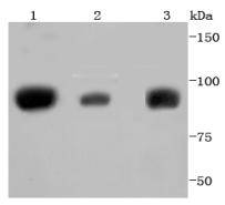
Fig1: Western blot analysis of STAT5A+B on different lysates using anti-STAT5A+B antibody at 1/1,000 dilution. Positive control: Lane 1: K562 Lane 2: Jurkat Lane 3: PC-12Application
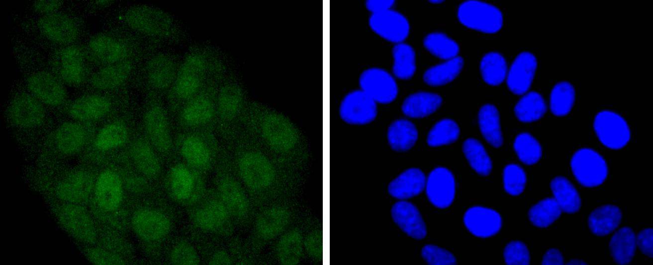
Fig2: ICC staining STAT5A+B in HepG2 cells (green). The nuclear counter stain is DAPI (blue). Cells were fixed in paraformaldehyde, permeabilised with 0.25% Triton X100/PBS.Application
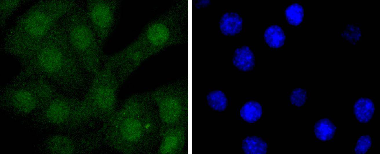
Fig3: ICC staining STAT5A+B in NIH/3T3 cells (green). The nuclear counter stain is DAPI (blue). Cells were fixed in paraformaldehyde, permeabilised with 0.25% Triton X100/PBS.Application
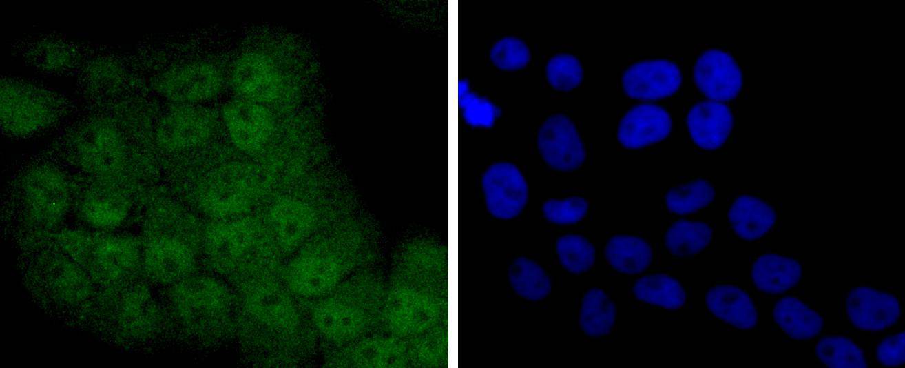
Fig4: ICC staining STAT5A+B in RH-35 cells (green). The nuclear counter stain is DAPI (blue). Cells were fixed in paraformaldehyde, permeabilised with 0.25% Triton X100/PBS.Application
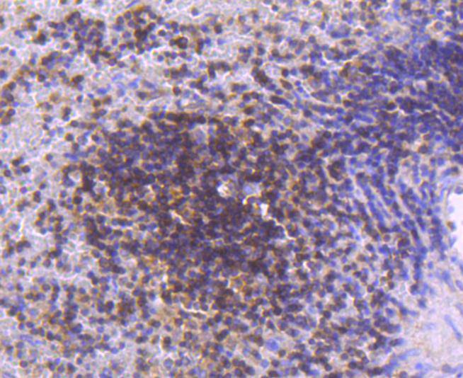
Fig5: Immunohistochemical analysis of paraffin-embedded human spleen tissue using anti-STAT5A+B antibody. Counter stained with hematoxylin.Application
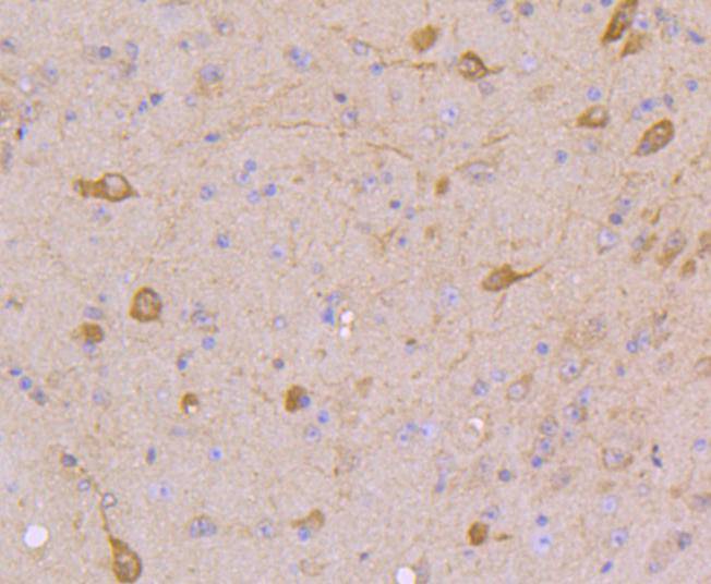
Fig6: Immunohistochemical analysis of paraffin-embedded mouse brain tissue using anti-STAT5A+B antibody. Counter stained with hematoxylin.Application
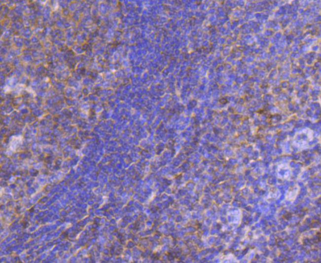
Fig7: Immunohistochemical analysis of paraffin-embedded human tonsil tissue using anti-STAT5A+B antibody. Counter stained with hematoxylin.Application
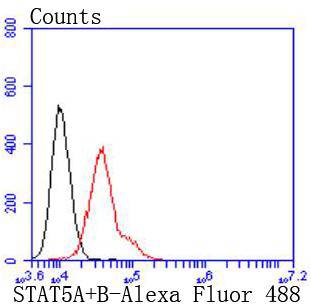
Fig8: Flow cytometric analysis of NIH/3T3 cells with STAT5A+B antibody at 1/50 dilution (red) compared with an unlabelled control (cells without incubation with primary antibody; black). Alexa Fluor 488-conjugated goat anti rabbit IgG was used as the secondary antibody.| Product Name | STAT 5A+B [JJ08-78] |
|---|---|
| Antibody Type | Primary Antibodies |
| Antigen Alias | Mammary gland factor (STAT5A) antibody MGF (STAT5A) antibody Signal transducer and activator of transcription 5A antibody Signal transducer and activator of transcription 5B antibody STA5A_HUMAN antibody Stat 5a antibody STAT 5b antibody STAT5A antibody Transcription factor STAT5B antibody |
| Product description | Stat5 (Signal Transducers and Activators of Transcription 5) is important in regulating T cell functions involving the receptors for Interleukin-2 (IL-2). IL-2 stimulates the rapid phosphorylation of both serine and tyrosine residues of Stat5a and Stat5b in human T lymphocytes and in several IL-2-responsive lymphocytic cell lines. IL-2 differentially induces serine phosphorylation of Stat5a and Stat5b on Ser726 and Ser731, respectively. Stat5b is preferentially phosphorylated and displays more protracted serine phosphorylation kinetics than Stat5a. Both the acid-rich region and the COOH terminus of IL-2Rb can independently mediate IL-2-induced Stat 5a/b serine phosphorylation, suggesting that Stat5a/b serine phosphorylation occurs at a postreceptor level. Stat5a is phosphorylated on Tyr694 in a prolactin-sensitive manner, whereas serine phosphorylation is constitutive. Activation of Stat5 by IL-2 may help govern the biological effects of IL-2 during the immune response. |
| Immunogen | recombinant protein |
| Clonality | Monoclonal |
|---|---|
| Isotype | IgG |
| Host Species | Recombinant rabbit |
| Tested Applications | WBICC/IFIHCIPFC |
| WB:1:1,000-1:2,000 ICC:1:50-1:200 IHC:1:50-1:200 FC:1:50-1:100 |
|
| Species Reactivity | HumanMouseRat |
| Concentration | 1mg/ml |
| Alternative Names | Mammary gland factor (STAT5A) antibody MGF (STAT5A) antibody Signal transducer and activator of transcription 5A antibody Signal transducer and activator of transcription 5B antibody STA5A_HUMAN antibody Stat 5a antibody STAT 5b antibody STAT5A antibody Transcription factor STAT5B antibody |
|---|---|
| Molecular Weight(MW) | 90 kDa |
| Cellular Localization | Cytoplasm, Nucleus. |
Application
Fig1: Western blot analysis of STAT5A+B on different lysates using anti-STAT5A+B antibody at 1/1,000 dilution. Positive control: Lane 1: K562 Lane 2: Jurkat Lane 3: PC-12
Application
Fig2: ICC staining STAT5A+B in HepG2 cells (green). The nuclear counter stain is DAPI (blue). Cells were fixed in paraformaldehyde, permeabilised with 0.25% Triton X100/PBS.
Application
Fig3: ICC staining STAT5A+B in NIH/3T3 cells (green). The nuclear counter stain is DAPI (blue). Cells were fixed in paraformaldehyde, permeabilised with 0.25% Triton X100/PBS.
Application
Fig4: ICC staining STAT5A+B in RH-35 cells (green). The nuclear counter stain is DAPI (blue). Cells were fixed in paraformaldehyde, permeabilised with 0.25% Triton X100/PBS.
Application
Fig5: Immunohistochemical analysis of paraffin-embedded human spleen tissue using anti-STAT5A+B antibody. Counter stained with hematoxylin.
Application
Fig6: Immunohistochemical analysis of paraffin-embedded mouse brain tissue using anti-STAT5A+B antibody. Counter stained with hematoxylin.
Application
Fig7: Immunohistochemical analysis of paraffin-embedded human tonsil tissue using anti-STAT5A+B antibody. Counter stained with hematoxylin.
Application
Fig8: Flow cytometric analysis of NIH/3T3 cells with STAT5A+B antibody at 1/50 dilution (red) compared with an unlabelled control (cells without incubation with primary antibody; black). Alexa Fluor 488-conjugated goat anti rabbit IgG was used as the secondary antibody.| Positive Control | K562, Jurkat, PC-12, HepG2, RH-35, NIH/3T3, human tonsil tissue, human spleen tissue, mouse brain tissue. |
|---|---|
| Application Notes | WB:1:1,000-1:2,000 ICC:1:50-1:200 IHC:1:50-1:200 FC:1:50-1:100 |
| Form | Liquid |
|---|---|
| Storage Instructions | Store at +4℃ after thawing. Aliquot store at -20℃ or -80℃. Avoid repeated freeze / thaw cycles. |
| Storage Buffer | 1*TBS (pH7.4), 1%BSA, 40%Glycerol. Preservative: 0.05% Sodium Azide. |


