Application
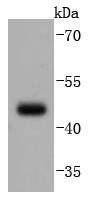
Fig1: Western blot analysis of PAX5 on Daudi cells lysates using anti-PAX5 antibody at 1/1,000 dilution.Application
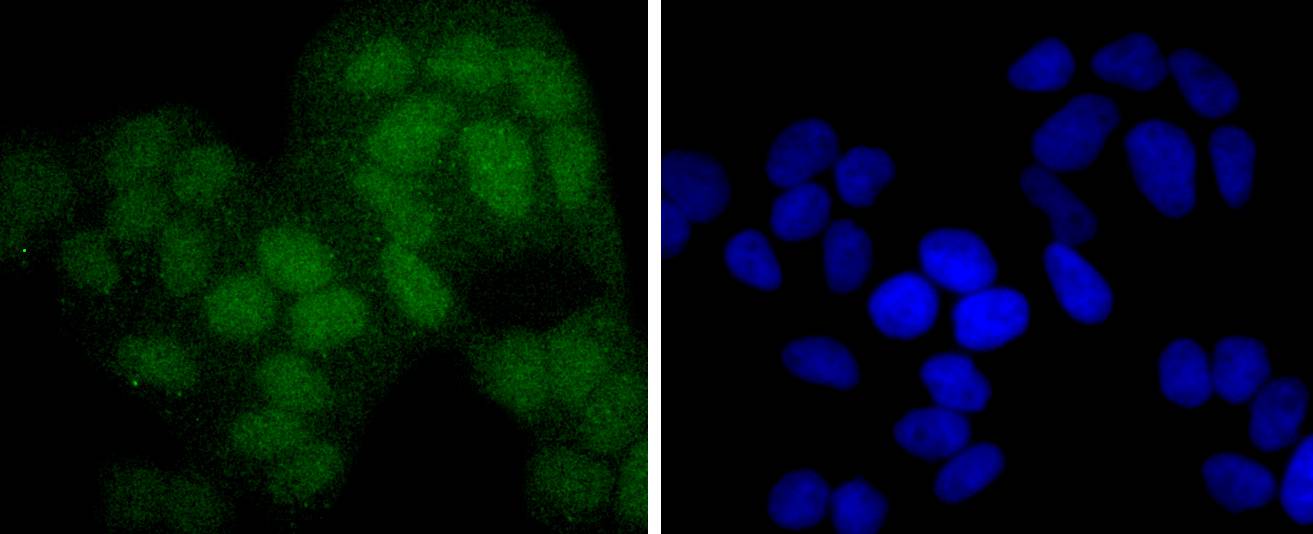
Fig2: ICC staining PAX5 in Hela cells (green). The nuclear counter stain is DAPI (blue). Cells were fixed in paraformaldehyde, permeabilised with 0.25% Triton X100/PBS.Application
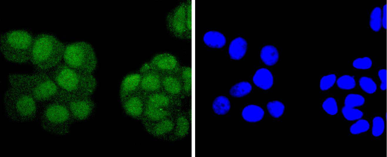
Fig3: ICC staining PAX5 in MCF-7 cells (green). The nuclear counter stain is DAPI (blue). Cells were fixed in paraformaldehyde, permeabilised with 0.25% Triton X100/PBS.Application
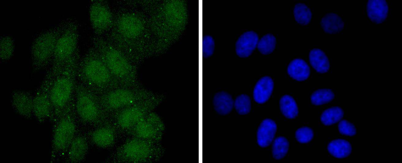
Fig4: ICC staining PAX5 in HepG2 cells (green). The nuclear counter stain is DAPI (blue). Cells were fixed in paraformaldehyde, permeabilised with 0.25% Triton X100/PBS.Application
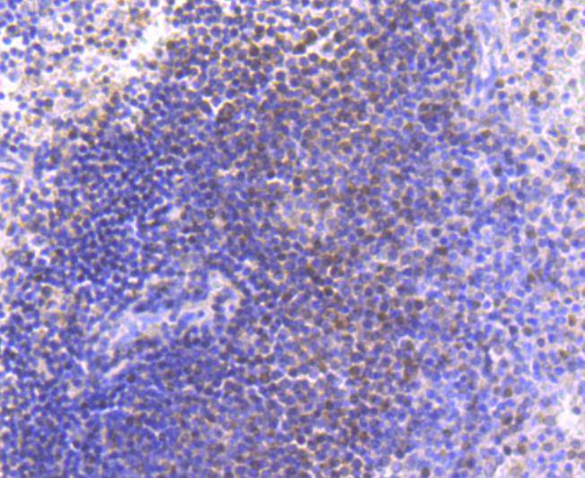
Fig5: Immunohistochemical analysis of paraffin-embedded human spleen tissue using anti-PAX5 antibody. Counter stained with hematoxylin.Application
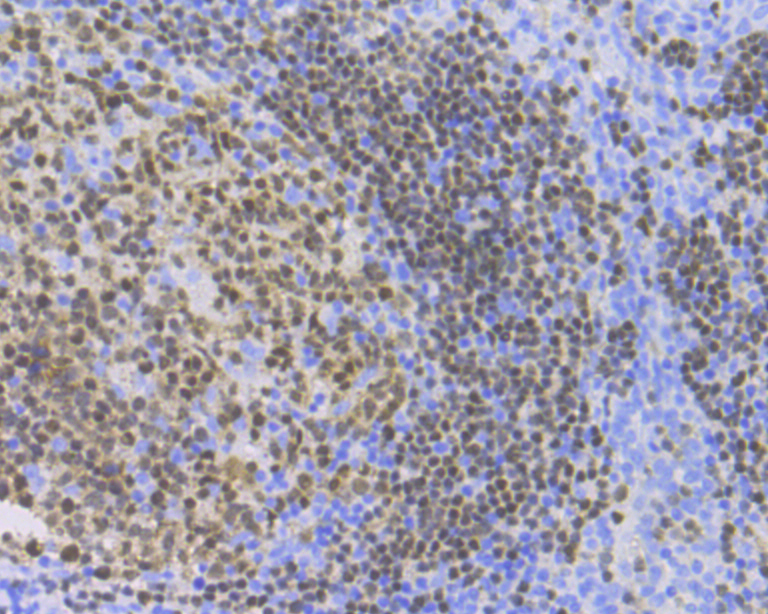
Fig6: Immunohistochemical analysis of paraffin-embedded human tonsil tissue using anti-PAX5 antibody. Counter stained with hematoxylin.Application
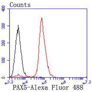
Fig7: Flow cytometric analysis of Raji cells with PAX5 antibody at 1/50 dilution (red) compared with an unlabelled control (cells without incubation with primary antibody; black). Alexa Fluor 488-conjugated goat anti rabbit IgG was used as the secondary antibody.| Product Name | PAX5 [JJ08-87] |
|---|---|
| Antibody Type | Primary Antibodies |
| Antigen Alias | B cell lineage specific activator antibody B cell lineage specific activator protein antibody B cell specific activator protein antibody B cell specific transcription factor antibody B-cell-specific transcription factor antibody BSAP antibody EBB-1 antibody KLP antibody Paired box 5 antibody Paired box gene 5 (B cell lineage specific activator protein) antibody Paired box gene 5 (B cell lineage specific activator) antibody Paired box gene 5 antibody Paired box homeotic gene 5 antibody Paired box protein Pax 5 antibody Paired box protein Pax-5 antibody Paired domain gene 5 antibody PAX 5 antibody PAX5 antibody PAX5_HUMAN antibody Transcription factor PAX 5 antibody |
| Product description | The Pax family of nuclear transcription factors is comprised of nine members that function during embryogenesis to regulate the temporal and position-dependent differentiation of cells. Pax family genes are also involved in a variety of signal transduction pathways in the adult organism. Mutations in Pax proteins have been linked to disease and cancer in humans. For example, the human PAX5 gene encodes a B cell lineage-specific protein, Pax-5, also designated B cell specific activator protein or BSAP, which is expressed in pro-B, pre-B and mature B lymphocytes but not in plasma cells. Pax-5 functions to regulate not only B cell development, but also influences the balance between immunoglobulin secretion and B cell proliferation. Overexpression of Pax-5 has been implicated in cellular transformation, and in the case of small lymphocytic lymphomas with plasmacytoid differentiation, a t(9;14)(p13;q32) translocation resulting in the deregulation of PAX5 gene expression has been detected. |
| Immunogen | recombinant protein |
| Clonality | Monoclonal |
|---|---|
| Isotype | IgG |
| Host Species | Recombinant rabbit |
| Tested Applications | WBICC/IFIHCFC |
| WB:1:1,000-1:2,000 ICC:1:50-1:200 IHC:1:50-1:200 FC:1:50-1:100 |
|
| Species Reactivity | HumanMouse |
| Concentration | 1mg/ml |
| Alternative Names | B cell lineage specific activator antibody B cell lineage specific activator protein antibody B cell specific activator protein antibody B cell specific transcription factor antibody B-cell-specific transcription factor antibody BSAP antibody EBB-1 antibody KLP antibody Paired box 5 antibody Paired box gene 5 (B cell lineage specific activator protein) antibody Paired box gene 5 (B cell lineage specific activator) antibody Paired box gene 5 antibody Paired box homeotic gene 5 antibody Paired box protein Pax 5 antibody Paired box protein Pax-5 antibody Paired domain gene 5 antibody PAX 5 antibody PAX5 antibody PAX5_HUMAN antibody Transcription factor PAX 5 antibody |
|---|---|
| Molecular Weight(MW) | 42 kDa |
| Cellular Localization | Nucleus. |
Application
Fig1: Western blot analysis of PAX5 on Daudi cells lysates using anti-PAX5 antibody at 1/1,000 dilution.
Application
Fig2: ICC staining PAX5 in Hela cells (green). The nuclear counter stain is DAPI (blue). Cells were fixed in paraformaldehyde, permeabilised with 0.25% Triton X100/PBS.
Application
Fig3: ICC staining PAX5 in MCF-7 cells (green). The nuclear counter stain is DAPI (blue). Cells were fixed in paraformaldehyde, permeabilised with 0.25% Triton X100/PBS.
Application
Fig4: ICC staining PAX5 in HepG2 cells (green). The nuclear counter stain is DAPI (blue). Cells were fixed in paraformaldehyde, permeabilised with 0.25% Triton X100/PBS.
Application
Fig5: Immunohistochemical analysis of paraffin-embedded human spleen tissue using anti-PAX5 antibody. Counter stained with hematoxylin.
Application
Fig6: Immunohistochemical analysis of paraffin-embedded human tonsil tissue using anti-PAX5 antibody. Counter stained with hematoxylin.
Application
Fig7: Flow cytometric analysis of Raji cells with PAX5 antibody at 1/50 dilution (red) compared with an unlabelled control (cells without incubation with primary antibody; black). Alexa Fluor 488-conjugated goat anti rabbit IgG was used as the secondary antibody.| Positive Control | Daudi, MCF-7, Hela, HepG2, human tonsil tissue, human spleen tissue. |
|---|---|
| Application Notes | WB:1:1,000-1:2,000 ICC:1:50-1:200 IHC:1:50-1:200 FC:1:50-1:100 |
| Form | Liquid |
|---|---|
| Storage Instructions | Store at +4℃ after thawing. Aliquot store at -20℃ or -80℃. Avoid repeated freeze / thaw cycles. |
| Storage Buffer | 1*TBS (pH7.4), 1%BSA, 40%Glycerol. Preservative: 0.05% Sodium Azide. |


