WB
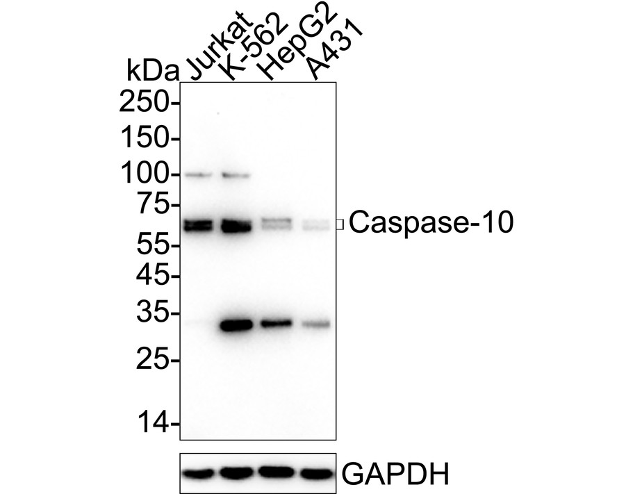
Western blot analysis of Caspase-10 on different lysates with Rabbit anti-Caspase-10 antibody at 1/2,000 dilution. Lane 1: Jurkat cell lysate, Lane 2: K-562 cell lysate, Lane 3: HepG2 cell lysate, Lane 4: A431 cell lysate, Lysates/proteins at 20 µg/Lane. Exposure time: 2 minutes; 4-20% SDS-PAGE gel. Proteins were transferred to a PVDF membrane and blocked with 5% NFDM/TBST for 1 hour at room temperature. The primary antibody at 1/2,000 dilution was used in 5% NFDM/TBST at 4℃ overnight. Goat Anti-Rabbit IgG - HRP Secondary Antibody at 1/50,000 dilution was used for 1 hour at room temperature.IHC
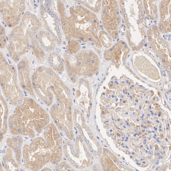
Immunohistochemical analysis of paraffin-embedded human kidney tissue with Rabbit anti-Caspase-10 antibody at 1/200 dilution. The section was pre-treated using heat mediated antigen retrieval with Tris-EDTA buffer (pH 9.0) for 20 minutes. The tissues were blocked in 1% BSA for 20 minutes at room temperature, washed with ddH2O and PBS, and then probed with the primary antibody at 1/200 dilution for 1 hour at room temperature. The detection was performed using an HRP conjugated compact polymer system. DAB was used as the chromogen. Tissues were counterstained with hematoxylin and mounted with DPX.ICC/IF
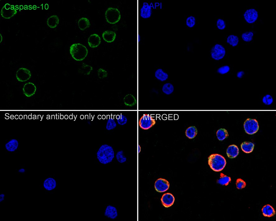
Immunocytochemistry analysis of Jurkat cells labeling Caspase-10 with Rabbit anti-Caspase-10 antibody at 1/100 dilution. Cells were fixed in 4% paraformaldehyde for 15 minutes at room temperature, permeabilized with 0.1% Triton X-100 in PBS for 15 minutes at room temperature, then blocked with 1% BSA in 10% negative goat serum for 1 hour at room temperature. Cells were then incubated with Rabbit anti-Caspase-10 antibody at 1/100 dilution in 1% BSA in PBST overnight at 4 ℃. Goat Anti-Rabbit IgG H&L (488) was used as the secondary antibody at 1/1,000 dilution. PBS instead of the primary antibody was used as the secondary antibody only control. Nuclear DNA was labelled in blue with DAPI. Beta tubulin (red) was stained at 1/100 dilution overnight at +4℃. Goat Anti-Mouse IgG H&L (594) was used as the secondary antibody at 1/1,000 dilution.FC
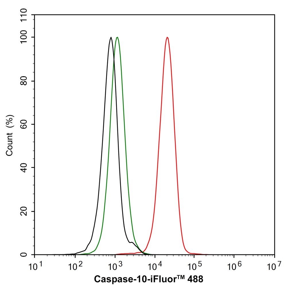
Flow cytometric analysis of Jurkat cells labeling Caspase-10. Cells were fixed and permeabilized. Then stained with the primary antibody (1/1,000) (red) compared with Rabbit IgG Isotype Control (green). After incubation of the primary antibody at +4℃ for an hour, the cells were stained with a iFluor™ 488 conjugate-Goat anti-Rabbit IgG Secondary antibody at 1/1,000 dilution for 30 minutes at +4℃. Unlabelled sample was used as a control (cells without incubation with primary antibody; black).IP
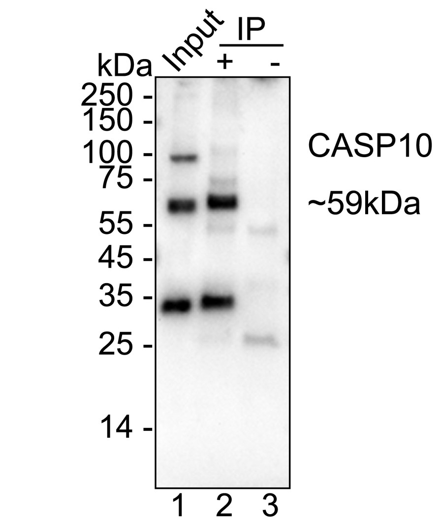
Caspase-10 was immunoprecipitated from 0.2 mg K-562 cell lysate with Rabbit anti-Caspase-10 antibody at 2 µg/10 µl beads. Western blot was performed from the immunoprecipitate using Rabbit anti-Caspase-10 antibody at 1/2,000 dilution. HRP Conjugated Anti-Rabbit IgG for IP Nano-secondary antibody at 1/5,000 dilution was used for 1 hour at room temperature. Lane 1: K-562 cell lysate (input), Lane 2: Rabbit anti-Caspase-10 antibody IP in K-562 cell lysate, Lane 3: Rabbit IgG instead of Rabbit anti-Caspase-10 antibody in K-562 cell lysate, Blocking/Dilution buffer: 5% NFDM/TBST, Exposure time: 1 minute 7 seconds.| Product Name | Caspase-10 Recombinant Rabbit Monoclonal Antibody |
|---|---|
| Antibody Type | Primary Antibodies |
| Product description | Caspase-10, also designated Mch4, is recruited to the native TRAIL and CD9 death-inducing signaling complexes (DISCs) by the FADD/Mort1 adaptor protein complex. Caspase-10 requires the assembly of the FADD and DISC complexes for its recruitment and cleavage-induced activation during CD95-induced apoptosis of activated T cells. The N-terminus of caspase-10 contains FADD-like death effector domains further indicating that it associates with FADD to induce apoptosis. Caspase-10 is not required for apoptosis induction and when overexpressed, cannot reverse defects in apoptosis induction caused by caspase-8 deficiency. Granzyme B cleaves procaspase-10 at an IXXD-A processing sequence to produce mature caspase-10. Mutations in the caspase-10 gene in the prodomain, p17 large protease subunit and p12 small protease subunit have been linked to a number of non-Hodgkin lymphomas in humans. |
| Immunogen | Recombinant protein within Human Caspase-10 aa 254-452 / 521. |
| Clonality | monoclonal |
|---|---|
| Isotype | IgG |
| Host Species | Recombinant rabbit |
| Tested Applications | FCICC/IFIHCIPWB |
| WB:1:1000-1:2000 IHC:1:50-1:200 ICC/IF:1:100 FC:1:1000 IP:1-2μg/sample |
|
| Species Reactivity | Human |
| Concentration | 1mg/ml |
| Purification | Protein A |
| Alternative Names | ALPS2 antibody Apoptotic protease Mch-4 antibody CASP 10 antibody CASP-10 antibody CASP10 antibody CASPA_HUMAN antibody Caspase 10 apoptosis related cysteine peptidase antibody Caspase-10 subunit p12 antibody FADD like ICE2 antibody Fas associated death domain protein antibody FAS-associated death domain protein interleukin-1B-converting enzyme 2 antibody FLICE 2 antibody FLICE2 antibody ICE like apoptotic protease 4 antibody ICE-like apoptotic protease 4 antibody Interleukin 1B converting enzyme 2 antibody MCH 4 antibody |
|---|---|
| Molecular Weight(MW) | 59 kDa |
| Cellular Localization | Cytosol. |
| SwissProt ID | Q92851 |
|---|
WB
Western blot analysis of Caspase-10 on different lysates with Rabbit anti-Caspase-10 antibody at 1/2,000 dilution. Lane 1: Jurkat cell lysate, Lane 2: K-562 cell lysate, Lane 3: HepG2 cell lysate, Lane 4: A431 cell lysate, Lysates/proteins at 20 µg/Lane. Exposure time: 2 minutes; 4-20% SDS-PAGE gel. Proteins were transferred to a PVDF membrane and blocked with 5% NFDM/TBST for 1 hour at room temperature. The primary antibody at 1/2,000 dilution was used in 5% NFDM/TBST at 4℃ overnight. Goat Anti-Rabbit IgG - HRP Secondary Antibody at 1/50,000 dilution was used for 1 hour at room temperature.
IHC
Immunohistochemical analysis of paraffin-embedded human kidney tissue with Rabbit anti-Caspase-10 antibody at 1/200 dilution. The section was pre-treated using heat mediated antigen retrieval with Tris-EDTA buffer (pH 9.0) for 20 minutes. The tissues were blocked in 1% BSA for 20 minutes at room temperature, washed with ddH2O and PBS, and then probed with the primary antibody at 1/200 dilution for 1 hour at room temperature. The detection was performed using an HRP conjugated compact polymer system. DAB was used as the chromogen. Tissues were counterstained with hematoxylin and mounted with DPX.
ICC/IF
Immunocytochemistry analysis of Jurkat cells labeling Caspase-10 with Rabbit anti-Caspase-10 antibody at 1/100 dilution. Cells were fixed in 4% paraformaldehyde for 15 minutes at room temperature, permeabilized with 0.1% Triton X-100 in PBS for 15 minutes at room temperature, then blocked with 1% BSA in 10% negative goat serum for 1 hour at room temperature. Cells were then incubated with Rabbit anti-Caspase-10 antibody at 1/100 dilution in 1% BSA in PBST overnight at 4 ℃. Goat Anti-Rabbit IgG H&L (488) was used as the secondary antibody at 1/1,000 dilution. PBS instead of the primary antibody was used as the secondary antibody only control. Nuclear DNA was labelled in blue with DAPI. Beta tubulin (red) was stained at 1/100 dilution overnight at +4℃. Goat Anti-Mouse IgG H&L (594) was used as the secondary antibody at 1/1,000 dilution.
FC
Flow cytometric analysis of Jurkat cells labeling Caspase-10. Cells were fixed and permeabilized. Then stained with the primary antibody (1/1,000) (red) compared with Rabbit IgG Isotype Control (green). After incubation of the primary antibody at +4℃ for an hour, the cells were stained with a iFluor™ 488 conjugate-Goat anti-Rabbit IgG Secondary antibody at 1/1,000 dilution for 30 minutes at +4℃. Unlabelled sample was used as a control (cells without incubation with primary antibody; black).
IP
Caspase-10 was immunoprecipitated from 0.2 mg K-562 cell lysate with Rabbit anti-Caspase-10 antibody at 2 µg/10 µl beads. Western blot was performed from the immunoprecipitate using Rabbit anti-Caspase-10 antibody at 1/2,000 dilution. HRP Conjugated Anti-Rabbit IgG for IP Nano-secondary antibody at 1/5,000 dilution was used for 1 hour at room temperature. Lane 1: K-562 cell lysate (input), Lane 2: Rabbit anti-Caspase-10 antibody IP in K-562 cell lysate, Lane 3: Rabbit IgG instead of Rabbit anti-Caspase-10 antibody in K-562 cell lysate, Blocking/Dilution buffer: 5% NFDM/TBST, Exposure time: 1 minute 7 seconds.| Application Notes | WB:1:1000-1:2000 IHC:1:50-1:200 ICC/IF:1:100 FC:1:1000 IP:1-2μg/sample |
|---|
| Form | Liquid |
|---|---|
| Storage Instructions | Store at +4℃ after thawing. Aliquot store at -20℃ or -80℃. Avoid repeated freeze / thaw cycles. |
| Storage Buffer | 1*TBS (pH7.4), 1%BSA, 40%Glycerol. Preservative: 0.05% Sodium Azide. |


