WB
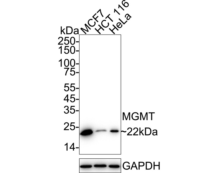
Western blot analysis of MGMT on different lysates with Rabbit anti-MGMT antibody at 1/5,000 dilution. Lane 1: MCF7 cell lysate, Lane 2: HCT 116 cell lysate, Lane 3: HeLa cell lysate, Lysates/proteins at 20 µg/Lane. Exposure time: 25 seconds; 4-20% SDS-PAGE gel. Proteins were transferred to a PVDF membrane and blocked with 5% NFDM/TBST for 1 hour at room temperature. The primary antibody at 1/5,000 dilution was used in 5% NFDM/TBST at 4℃ overnight. Goat Anti-Rabbit IgG - HRP Secondary Antibody at 1/50,000 dilution was used for 1 hour at room temperature.WB
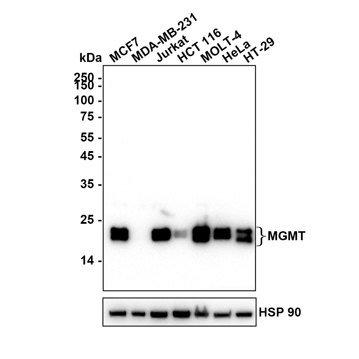
Western blot analysis of MGMT on different lysates with Rabbit anti-MGMT antibody at 1/5,000 dilution. Lane 1: MCF7 (Human breast cancer cell) cell lysate, Lane 2: MDA-MB-231 (Human breast cancer cell) cell lysate, Lane 3: Jurkat (Human T-lymphoblastic cells) cell lysate, Lane 4: HCT 116 (Human colon cancer cell) cell lysate, Lane 5: MOLT-4 (Human acute T-Cell leukemia cell) cell lysate, Lane 6: HeLa (Human cervical adenocarcinoma cell) cell lysate, Lane 7: HT-29 (Human colorectal adenocarcinoma cell) cell lysate, Lysates/proteins at 15 µg/Lane. Exposure time: 25 seconds; MDA-MB-231 exhibits low MGMT expression (PMID: 26420420). Blocking: 5% NFDM/TBST, 1 hour at room temperature, Primary antibody:1/5,000 in primary antibody dilution buffer, overnight at 4 ℃ Secondary antibody: Goat anti-Rabbit IgG-HRP, 1/50,000 in 5% NFDM/TBST, 1 hour at room temperature.ICC/IF
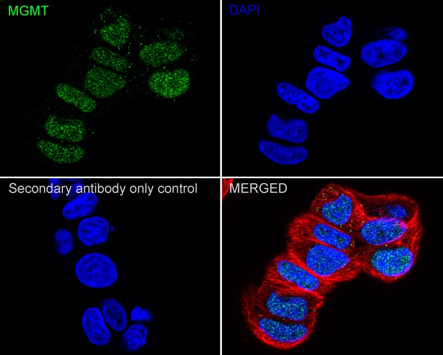
Immunocytochemistry analysis of MCF7 cells labeling MGMT with Rabbit anti-MGMT antibody at 1/100 dilution. Cells were fixed in 4% paraformaldehyde for 15 minutes at room temperature, permeabilized with 0.1% Triton X-100 in PBS for 15 minutes at room temperature, then blocked with 1% BSA in 10% negative goat serum for 1 hour at room temperature. Cells were then incubated with Rabbit anti-MGMT antibody at 1/100 dilution in 1% BSA in PBST overnight at 4 ℃. Goat Anti-Rabbit IgG H&L (488) was used as the secondary antibody at 1/1,000 dilution. PBS instead of the primary antibody was used as the secondary antibody only control. Nuclear DNA was labelled in blue with DAPI. Beta tubulin (red) was stained at 1/100 dilution overnight at +4℃. Goat Anti-Mouse IgG H&L (594) was used as the secondary antibody at 1/1,000 dilution.FC
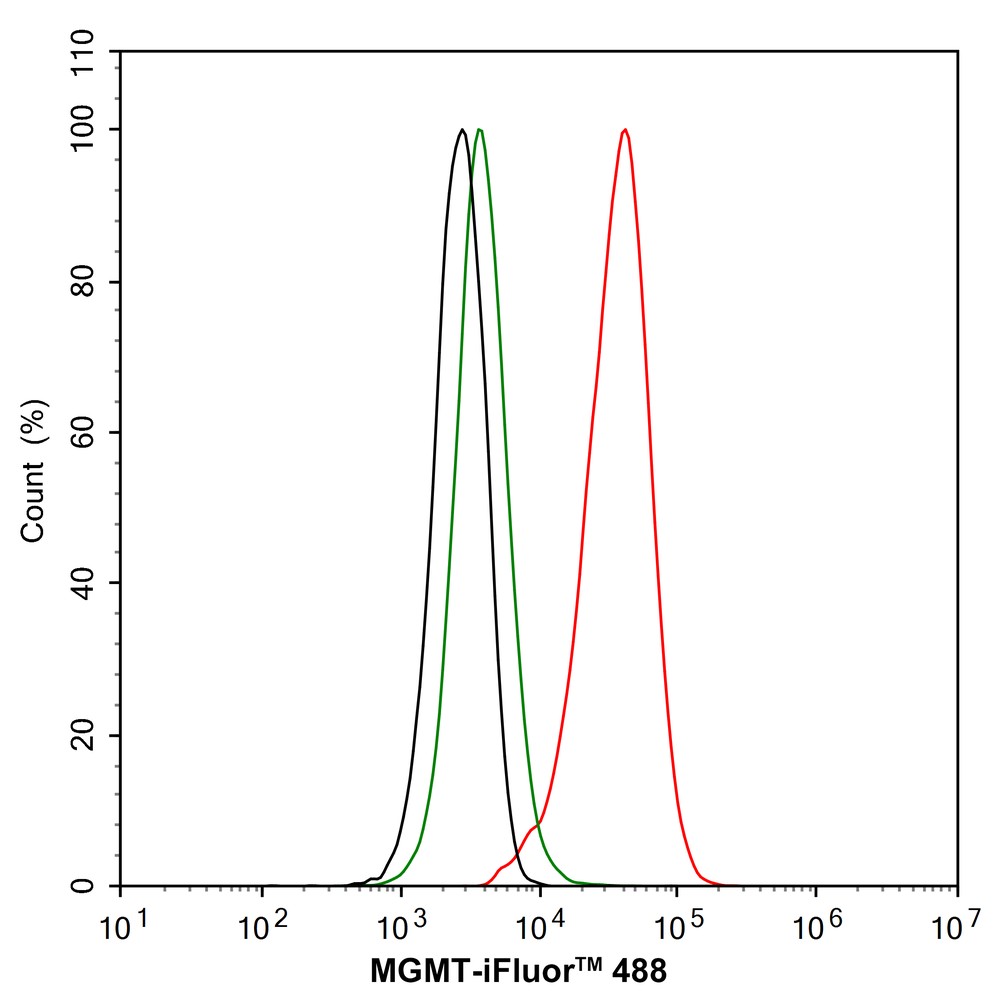
Flow cytometric analysis of MCF7 cells labeling MGMT. Cells were fixed and permeabilized. Then stained with the primary antibody (1/1,000) (red) compared with Rabbit IgG Isotype Control (green). After incubation of the primary antibody at +4℃ for an hour, the cells were stained with a 488 conjugate-Goat anti-Rabbit IgG Secondary antibody at 1/1,000 dilution for 30 minutes at +4℃. Unlabelled sample was used as a control (cells without incubation with primary antibody; black).IHC
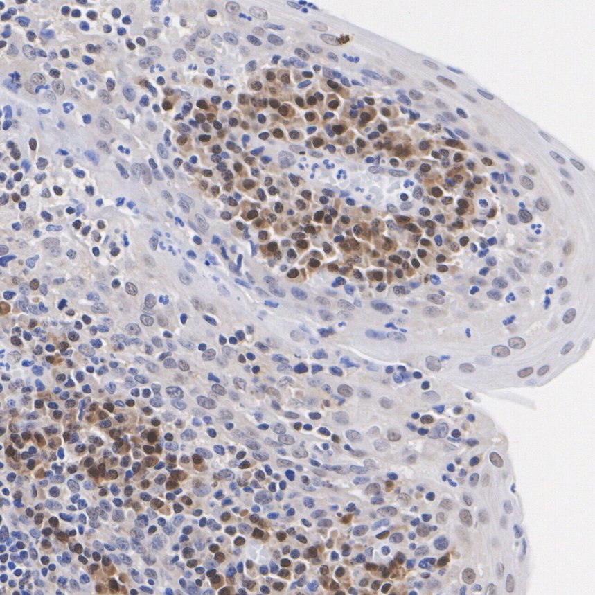
Species: Human, Tissue: Tonsil, Sample: Paraffin-embedded section, Antigen retrieval: Heat-mediated, Tris-EDTA buffer (pH 9.0), 20 minutes at 95℃. Wash buffer: 1× TBST, Endogenous peroxidase blocking: 3% H₂O₂, 10 minutes at room temperature. Blocking: 1% BSA + 10% normal goat serum, 10 minutes at room temperature. Primary antibody: 1/200, 1 hour at room temperature. Secondary antibody:20 minutes at room temperature.IHC
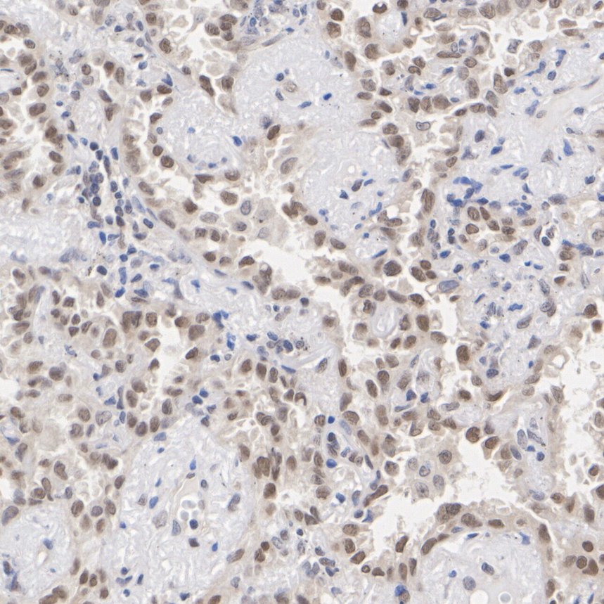
Species: Human, Tissue: Lung carcinoma, Sample: Paraffin-embedded section, Antigen retrieval: Heat-mediated, Tris-EDTA buffer (pH 9.0), 20 minutes at 95℃. Wash buffer: 1× TBST Endogenous peroxidase blocking: 3% H₂O₂, 10 minutes at room temperature. Blocking: 1% BSA + 10% normal goat serum, 10 minutes at room temperature. Primary antibody: 1/50, 1 hour at room temperature. Secondary antibody: 20 minutes at room temperature.| Product Name | MGMT Recombinant Rabbit Monoclonal Antibody |
|---|---|
| Antibody Type | Primary Antibodies |
| Product description | MGMT (O6-methylguanine-DNA methyltransferase) is transcriptionally activated in response to DNA damage and functions to repair mutagenic and cytotoxic O6-alkylguanine lesions caused by carcinogens or cytostatic drugs. MGMT induction by ionising radiation does not occur in p53-deficient mice, suggesting that MGMT induction may require p53. Similarly, MGMT mRNA and protein were shown to be inducible by ionising radiation, only in cell lines that express functional p53, and not in cell lines that do not express wild type p53. In contrast, high MGMT activity was associated with the presence of mutant p53, in a study of oral cancer cell lines. Similarly, MGMT activity was significantly lower in ovarian tumors with wildtype p53 than in tumors with mutant p53, supporting the view that wildtype p53 down-regulates the basal MGMT promoter. |
| Immunogen | Recombinant protein within Human MGMT aa 1-140 / 207. |
| Clonality | Monoclonal |
|---|---|
| Isotype | IgG |
| Host Species | Recombinant rabbit |
| Tested Applications | FCICC/IFIHCWB |
| WB:1:1000-1:5000 IHC:1:50-1:200 ICC/IF:1:100 FC:1:1000 |
|
| Species Reactivity | Human |
| Concentration | 1mg/ml |
| Purification | Protein A |
| Alternative Names | 6 O methylguanine DNA methyltransferase antibody 6-O-methylguanine-DNA methyltransferase antibody Agat antibody AGT antibody AI267024 antibody EC 2.1.1.63 antibody Methylated DNA protein cysteine methyltransferase antibody Methylated-DNAprotein-cysteine methyltransferase antibody Methylguanine DNA methyltransferase antibody MGC107020 antibody MGMT antibody MGMT_HUMAN antibody O 6 methylguanine DNA alkyltransferase antibody O 6 methylguanine DNA methyltransferase antibody O-6-methylguanine-DNA methyltransferase antibody O-6-methylguanine-DNA-alkyltransferase antibody |
|---|---|
| Molecular Weight(MW) | 22 kDa |
| Cellular Localization | Nucleus. |
| SwissProt ID | P16455 |
|---|
WB
Western blot analysis of MGMT on different lysates with Rabbit anti-MGMT antibody at 1/5,000 dilution. Lane 1: MCF7 cell lysate, Lane 2: HCT 116 cell lysate, Lane 3: HeLa cell lysate, Lysates/proteins at 20 µg/Lane. Exposure time: 25 seconds; 4-20% SDS-PAGE gel. Proteins were transferred to a PVDF membrane and blocked with 5% NFDM/TBST for 1 hour at room temperature. The primary antibody at 1/5,000 dilution was used in 5% NFDM/TBST at 4℃ overnight. Goat Anti-Rabbit IgG - HRP Secondary Antibody at 1/50,000 dilution was used for 1 hour at room temperature.
WB
Western blot analysis of MGMT on different lysates with Rabbit anti-MGMT antibody at 1/5,000 dilution. Lane 1: MCF7 (Human breast cancer cell) cell lysate, Lane 2: MDA-MB-231 (Human breast cancer cell) cell lysate, Lane 3: Jurkat (Human T-lymphoblastic cells) cell lysate, Lane 4: HCT 116 (Human colon cancer cell) cell lysate, Lane 5: MOLT-4 (Human acute T-Cell leukemia cell) cell lysate, Lane 6: HeLa (Human cervical adenocarcinoma cell) cell lysate, Lane 7: HT-29 (Human colorectal adenocarcinoma cell) cell lysate, Lysates/proteins at 15 µg/Lane. Exposure time: 25 seconds; MDA-MB-231 exhibits low MGMT expression (PMID: 26420420). Blocking: 5% NFDM/TBST, 1 hour at room temperature, Primary antibody:1/5,000 in primary antibody dilution buffer, overnight at 4 ℃ Secondary antibody: Goat anti-Rabbit IgG-HRP, 1/50,000 in 5% NFDM/TBST, 1 hour at room temperature.
ICC/IF
Immunocytochemistry analysis of MCF7 cells labeling MGMT with Rabbit anti-MGMT antibody at 1/100 dilution. Cells were fixed in 4% paraformaldehyde for 15 minutes at room temperature, permeabilized with 0.1% Triton X-100 in PBS for 15 minutes at room temperature, then blocked with 1% BSA in 10% negative goat serum for 1 hour at room temperature. Cells were then incubated with Rabbit anti-MGMT antibody at 1/100 dilution in 1% BSA in PBST overnight at 4 ℃. Goat Anti-Rabbit IgG H&L (488) was used as the secondary antibody at 1/1,000 dilution. PBS instead of the primary antibody was used as the secondary antibody only control. Nuclear DNA was labelled in blue with DAPI. Beta tubulin (red) was stained at 1/100 dilution overnight at +4℃. Goat Anti-Mouse IgG H&L (594) was used as the secondary antibody at 1/1,000 dilution.
FC
Flow cytometric analysis of MCF7 cells labeling MGMT. Cells were fixed and permeabilized. Then stained with the primary antibody (1/1,000) (red) compared with Rabbit IgG Isotype Control (green). After incubation of the primary antibody at +4℃ for an hour, the cells were stained with a 488 conjugate-Goat anti-Rabbit IgG Secondary antibody at 1/1,000 dilution for 30 minutes at +4℃. Unlabelled sample was used as a control (cells without incubation with primary antibody; black).
IHC
Species: Human, Tissue: Tonsil, Sample: Paraffin-embedded section, Antigen retrieval: Heat-mediated, Tris-EDTA buffer (pH 9.0), 20 minutes at 95℃. Wash buffer: 1× TBST, Endogenous peroxidase blocking: 3% H₂O₂, 10 minutes at room temperature. Blocking: 1% BSA + 10% normal goat serum, 10 minutes at room temperature. Primary antibody: 1/200, 1 hour at room temperature. Secondary antibody:20 minutes at room temperature.
IHC
Species: Human, Tissue: Lung carcinoma, Sample: Paraffin-embedded section, Antigen retrieval: Heat-mediated, Tris-EDTA buffer (pH 9.0), 20 minutes at 95℃. Wash buffer: 1× TBST Endogenous peroxidase blocking: 3% H₂O₂, 10 minutes at room temperature. Blocking: 1% BSA + 10% normal goat serum, 10 minutes at room temperature. Primary antibody: 1/50, 1 hour at room temperature. Secondary antibody: 20 minutes at room temperature.| Application Notes | WB:1:1000-1:5000 IHC:1:50-1:200 ICC/IF:1:100 FC:1:1000 |
|---|
| Form | Liquid |
|---|---|
| Storage Instructions | Store at +4℃ after thawing. Aliquot store at -20℃ or -80℃. Avoid repeated freeze / thaw cycles. |
| Storage Buffer | 1*TBS (pH7.4), 1%BSA, 40%Glycerol. Preservative: 0.05% Sodium Azide. |


