Application
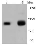
Fig1: Western blot analysis of KMT6/EZH2 on different lysates using anti-KMT6/EZH2 antibody at 1/1,000 dilution. Positive control: Lane 1: 293T Lane 2: 293Application
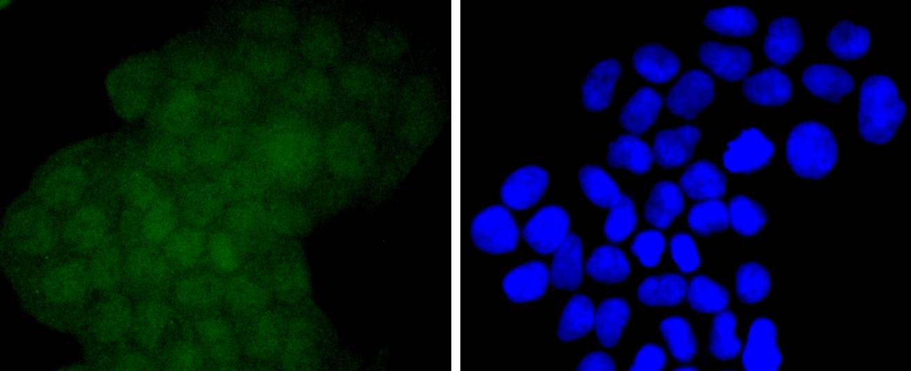
Fig2: ICC staining KMT6/EZH2 in Hela cells (green). The nuclear counter stain is DAPI (blue). Cells were fixed in paraformaldehyde, permeabilised with 0.25% Triton X100/PBS.Application
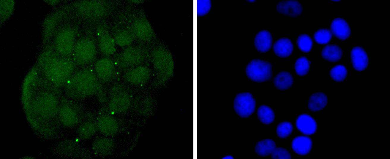
Fig3: ICC staining KMT6/EZH2 in MCF-7 cells (green). The nuclear counter stain is DAPI (blue). Cells were fixed in paraformaldehyde, permeabilised with 0.25% Triton X100/PBS.Application
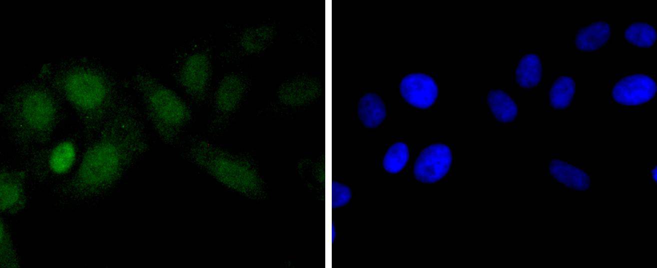
Fig4: ICC staining KMT6/EZH2 in HepG2 cells (green). The nuclear counter stain is DAPI (blue). Cells were fixed in paraformaldehyde, permeabilised with 0.25% Triton X100/PBS.Application
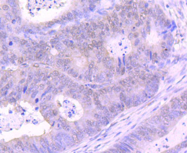
Fig5: Immunohistochemical analysis of paraffin-embedded human colon cancer tissue using anti-KMT6/EZH2 antibody. Counter stained with hematoxylin.Application
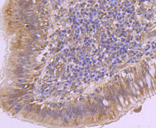
Fig6: Immunohistochemical analysis of paraffin-embedded human lung cancer tissue using anti-KMT6/EZH2 antibody. Counter stained with hematoxylin.Application
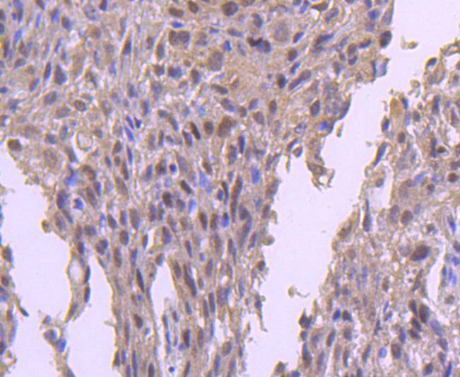
Fig7: Immunohistochemical analysis of paraffin-embedded human breast carcinoma tissue using anti-KMT6/EZH2 antibody. Counter stained with hematoxylin.| Product Name | KMT6/EZH2 [JJ089-9] |
|---|---|
| Antibody Type | Primary Antibodies |
| Antigen Alias | Enhancer of zeste 2 antibody enhancer of zeste 2 polycomb repressive complex 2 subunit antibody Enhancer of zeste homolog 2 (Drosophila) antibody Enhancer of zeste homolog 2 antibody Enhancer of zeste, Drosophila, homolog 2 antibody ENX 1 antibody Enx 1h antibody ENX-1 antibody ENX1 antibody Enx1h antibody EZH 2 antibody EZH1 antibody EZH2 antibody EZH2_HUMAN antibody EZH2b antibody Histone-lysine N-methyltransferase EZH2 antibody KMT 6 antibody KMT6 antibody KMT6A antibody Lysine N-methyltransferase 6 antibody MGC9169 antibody WVS antibody WVS2 antibody |
| Product description | In Drosophila, the Polycomb (PcG) gene family encodes chromatin proteins that are required for the repression of homeotic loci in embryonic development. PcG proteins work in conjunction with the trithorax-group (trxG), which activate homeobox gene expression during embryonic development. ENX-1, a mammalian homolog of the Drosophila gene enhancer of zeste, is a PcG protein that is ubiquitously expressed during early embryogenesis and becomes restricted to the central and peripheral nervous systems and sites of fetal hematopoiesis during later development. In the adult, ENX-1 is restricted to specific sites, including spleen, testis and placenta. The gene encoding human ENX-1 transcribes a 746 amino acid polypeptide which contains a trithorax-like domain and a DNA-binding motif. ENX-1 interacts with the proto-oncogene product Vav and is thought to be involved in the proliferation of normal and malignant hematopoietic cells. By altering the regulation of target genes, ENX-1 may also contribute to certain phenotypes of Down syndrome. |
| Immunogen | recombinant protein |
| Clonality | Monoclonal |
|---|---|
| Isotype | IgG |
| Host Species | Recombinant rabbit |
| Tested Applications | WBICC/IFIHC |
| WB:1:1,000-1:2,000 ICC:1:50-1:200 IHC:1:50-1:200 |
|
| Species Reactivity | Human |
| Concentration | 1mg/ml |
| Alternative Names | Enhancer of zeste 2 antibody
enhancer of zeste 2 polycomb repressive complex 2 subunit antibody
Enhancer of zeste homolog 2 (Drosophila) antibody
Enhancer of zeste homolog 2 antibody
Enhancer of zeste Drosophila homolog 2 antibody ENX 1 antibody Enx 1h antibody ENX-1 antibody ENX1 antibody Enx1h antibody EZH 2 antibody EZH1 antibody EZH2 antibody EZH2_HUMAN antibody EZH2b antibody Histone-lysine N-methyltransferase EZH2 antibody KMT 6 antibody KMT6 antibody KMT6A antibody Lysine N-methyltransferase 6 antibody MGC9169 antibody WVS antibody WVS2 antibody |
|---|---|
| Molecular Weight(MW) | 86 kDa |
| Cellular Localization | Nucleus. |
| SwissProt ID | Q15910 |
|---|
Application
Fig1: Western blot analysis of KMT6/EZH2 on different lysates using anti-KMT6/EZH2 antibody at 1/1,000 dilution. Positive control: Lane 1: 293T Lane 2: 293
Application
Fig2: ICC staining KMT6/EZH2 in Hela cells (green). The nuclear counter stain is DAPI (blue). Cells were fixed in paraformaldehyde, permeabilised with 0.25% Triton X100/PBS.
Application
Fig3: ICC staining KMT6/EZH2 in MCF-7 cells (green). The nuclear counter stain is DAPI (blue). Cells were fixed in paraformaldehyde, permeabilised with 0.25% Triton X100/PBS.
Application
Fig4: ICC staining KMT6/EZH2 in HepG2 cells (green). The nuclear counter stain is DAPI (blue). Cells were fixed in paraformaldehyde, permeabilised with 0.25% Triton X100/PBS.
Application
Fig5: Immunohistochemical analysis of paraffin-embedded human colon cancer tissue using anti-KMT6/EZH2 antibody. Counter stained with hematoxylin.
Application
Fig6: Immunohistochemical analysis of paraffin-embedded human lung cancer tissue using anti-KMT6/EZH2 antibody. Counter stained with hematoxylin.
Application
Fig7: Immunohistochemical analysis of paraffin-embedded human breast carcinoma tissue using anti-KMT6/EZH2 antibody. Counter stained with hematoxylin.| Positive Control | 293, MCF-7, Hela, HepG2, 293T, human lung cancer tissue, human colon cancer tissue, human breast carcinoma tissue. |
|---|---|
| Application Notes | WB:1:1,000-1:2,000 ICC:1:50-1:200 IHC:1:50-1:200 |
| Form | Liquid |
|---|---|
| Storage Instructions | Store at +4℃ after thawing. Aliquot store at -20℃ or -80℃. Avoid repeated freeze / thaw cycles. |
| Storage Buffer | 1*TBS (pH7.4), 1%BSA, 40%Glycerol. Preservative: 0.05% Sodium Azide. |


