Application
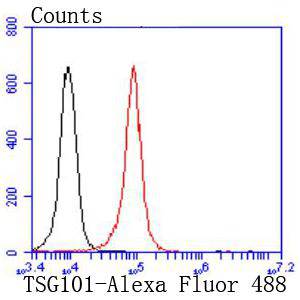
Fig9: Flow cytometric analysis of K562 cells with TSG101 antibody at 1/50 dilution (red) compared with an unlabelled control (cells without incubation with primary antibody; black). Alexa Fluor 488-conjugated goat anti rabbit IgG was used as the secondary antibody.Application
Fig9: Flow cytometric analysis of K562 cells with TSG101 antibody at 1/50 dilution (red) compared with an unlabelled control (cells without incubation with primary antibody; black). Alexa Fluor 488-conjugated goat anti rabbit IgG was used as the secondary antibody.Application
Fig9: Flow cytometric analysis of K562 cells with TSG101 antibody at 1/50 dilution (red) compared with an unlabelled control (cells without incubation with primary antibody; black). Alexa Fluor 488-conjugated goat anti rabbit IgG was used as the secondary antibody.Application
Fig9: Flow cytometric analysis of K562 cells with TSG101 antibody at 1/50 dilution (red) compared with an unlabelled control (cells without incubation with primary antibody; black). Alexa Fluor 488-conjugated goat anti rabbit IgG was used as the secondary antibody.Application
Fig9: Flow cytometric analysis of K562 cells with TSG101 antibody at 1/50 dilution (red) compared with an unlabelled control (cells without incubation with primary antibody; black). Alexa Fluor 488-conjugated goat anti rabbit IgG was used as the secondary antibody.Application
Fig9: Flow cytometric analysis of K562 cells with TSG101 antibody at 1/50 dilution (red) compared with an unlabelled control (cells without incubation with primary antibody; black). Alexa Fluor 488-conjugated goat anti rabbit IgG was used as the secondary antibody.Application
Fig9: Flow cytometric analysis of K562 cells with TSG101 antibody at 1/50 dilution (red) compared with an unlabelled control (cells without incubation with primary antibody; black). Alexa Fluor 488-conjugated goat anti rabbit IgG was used as the secondary antibody.Application
Fig9: Flow cytometric analysis of K562 cells with TSG101 antibody at 1/50 dilution (red) compared with an unlabelled control (cells without incubation with primary antibody; black). Alexa Fluor 488-conjugated goat anti rabbit IgG was used as the secondary antibody.Application
Fig9: Flow cytometric analysis of K562 cells with TSG101 antibody at 1/50 dilution (red) compared with an unlabelled control (cells without incubation with primary antibody; black). Alexa Fluor 488-conjugated goat anti rabbit IgG was used as the secondary antibody.| Product Name | TSG101 [JJ0900] |
|---|---|
| Antibody Type | Primary Antibodies |
| Antigen Alias | ESCRT I complex subunit TSG101 antibody ESCRT-I complex subunit TSG101 antibody TS101_HUMAN antibody TSG 10 antibody TSG 101 antibody TSG10 antibody Tsg101 antibody Tumor susceptibility gene 10 antibody Tumor susceptibility gene 101 antibody Tumor susceptibility gene 101 protein antibody Tumor susceptibility protein antibody Tumor susceptibility protein isoform 3 antibody VPS 23 antibody VPS23 antibody |
| Product description | The transformation of a normal cell to one that is malignant can result from mutations in genes that encode proteins with key regulatory functions. Examples include the retinoblastoma gene product (Rb p110), p53, VHL and APC. Using a novel cloning strategy that allows the isolation of previously uncharacterized genes encoding selectable recessive phenotypes, an additional tumor suppressor gene has been identified. This gene, termed tsg 101 for tumor susceptibility gene 101, encodes a stathmin binding domain protein. When expression of this growth inhibitory gene is blocked in NIH/3T3 cells using antisense mRNA, the cells exhibit a transformed phenotype and are tumorigenic in SL6 mice. |
| Immunogen | recombinant protein |
| Clonality | Monoclonal |
|---|---|
| Isotype | IgG |
| Host Species | Recombinant rabbit |
| Tested Applications | WBICC/IFIHCFC |
| WB:1:1,000-1:2,000 ICC:1:100-1:500 IHC:1:50-1:200 FC:1:50-1:100 |
|
| Species Reactivity | HumanMouseRat |
| Concentration | 1mg/ml |
| Alternative Names | ESCRT I complex subunit TSG101 antibody ESCRT-I complex subunit TSG101 antibody TS101_HUMAN antibody TSG 10 antibody TSG 101 antibody TSG10 antibody Tsg101 antibody Tumor susceptibility gene 10 antibody Tumor susceptibility gene 101 antibody Tumor susceptibility gene 101 protein antibody Tumor susceptibility protein antibody Tumor susceptibility protein isoform 3 antibody VPS 23 antibody VPS23 antibody |
|---|---|
| Molecular Weight(MW) | 44 kDa |
| Cellular Localization | Cytoplasm, Nucleus, Membrane, Late endosome membrane. |
Application
Fig9: Flow cytometric analysis of K562 cells with TSG101 antibody at 1/50 dilution (red) compared with an unlabelled control (cells without incubation with primary antibody; black). Alexa Fluor 488-conjugated goat anti rabbit IgG was used as the secondary antibody.
Application
Fig9: Flow cytometric analysis of K562 cells with TSG101 antibody at 1/50 dilution (red) compared with an unlabelled control (cells without incubation with primary antibody; black). Alexa Fluor 488-conjugated goat anti rabbit IgG was used as the secondary antibody.
Application
Fig9: Flow cytometric analysis of K562 cells with TSG101 antibody at 1/50 dilution (red) compared with an unlabelled control (cells without incubation with primary antibody; black). Alexa Fluor 488-conjugated goat anti rabbit IgG was used as the secondary antibody.
Application
Fig9: Flow cytometric analysis of K562 cells with TSG101 antibody at 1/50 dilution (red) compared with an unlabelled control (cells without incubation with primary antibody; black). Alexa Fluor 488-conjugated goat anti rabbit IgG was used as the secondary antibody.
Application
Fig9: Flow cytometric analysis of K562 cells with TSG101 antibody at 1/50 dilution (red) compared with an unlabelled control (cells without incubation with primary antibody; black). Alexa Fluor 488-conjugated goat anti rabbit IgG was used as the secondary antibody.
Application
Fig9: Flow cytometric analysis of K562 cells with TSG101 antibody at 1/50 dilution (red) compared with an unlabelled control (cells without incubation with primary antibody; black). Alexa Fluor 488-conjugated goat anti rabbit IgG was used as the secondary antibody.
Application
Fig9: Flow cytometric analysis of K562 cells with TSG101 antibody at 1/50 dilution (red) compared with an unlabelled control (cells without incubation with primary antibody; black). Alexa Fluor 488-conjugated goat anti rabbit IgG was used as the secondary antibody.
Application
Fig9: Flow cytometric analysis of K562 cells with TSG101 antibody at 1/50 dilution (red) compared with an unlabelled control (cells without incubation with primary antibody; black). Alexa Fluor 488-conjugated goat anti rabbit IgG was used as the secondary antibody.
Application
Fig9: Flow cytometric analysis of K562 cells with TSG101 antibody at 1/50 dilution (red) compared with an unlabelled control (cells without incubation with primary antibody; black). Alexa Fluor 488-conjugated goat anti rabbit IgG was used as the secondary antibody.| Positive Control | A431, NIH/3T3, Hela, SW480, K562, Jurkat, human colon cancer tissue, human kidney tissue, mouse colon tissue, mouse kidney tissue. |
|---|---|
| Application Notes | WB:1:1,000-1:2,000 ICC:1:100-1:500 IHC:1:50-1:200 FC:1:50-1:100 |
| Form | Liquid |
|---|---|
| Storage Instructions | Store at +4℃ after thawing. Aliquot store at -20℃ or -80℃. Avoid repeated freeze / thaw cycles. |
| Storage Buffer | 1*TBS (pH7.4), 1%BSA, 40%Glycerol. Preservative: 0.05% Sodium Azide. |


