Application
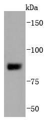
Fig1: Western blot analysis of SUZ12 on MCF-7 cells lysates using anti-SUZ12 antibody at 1/1,000 dilution.Application
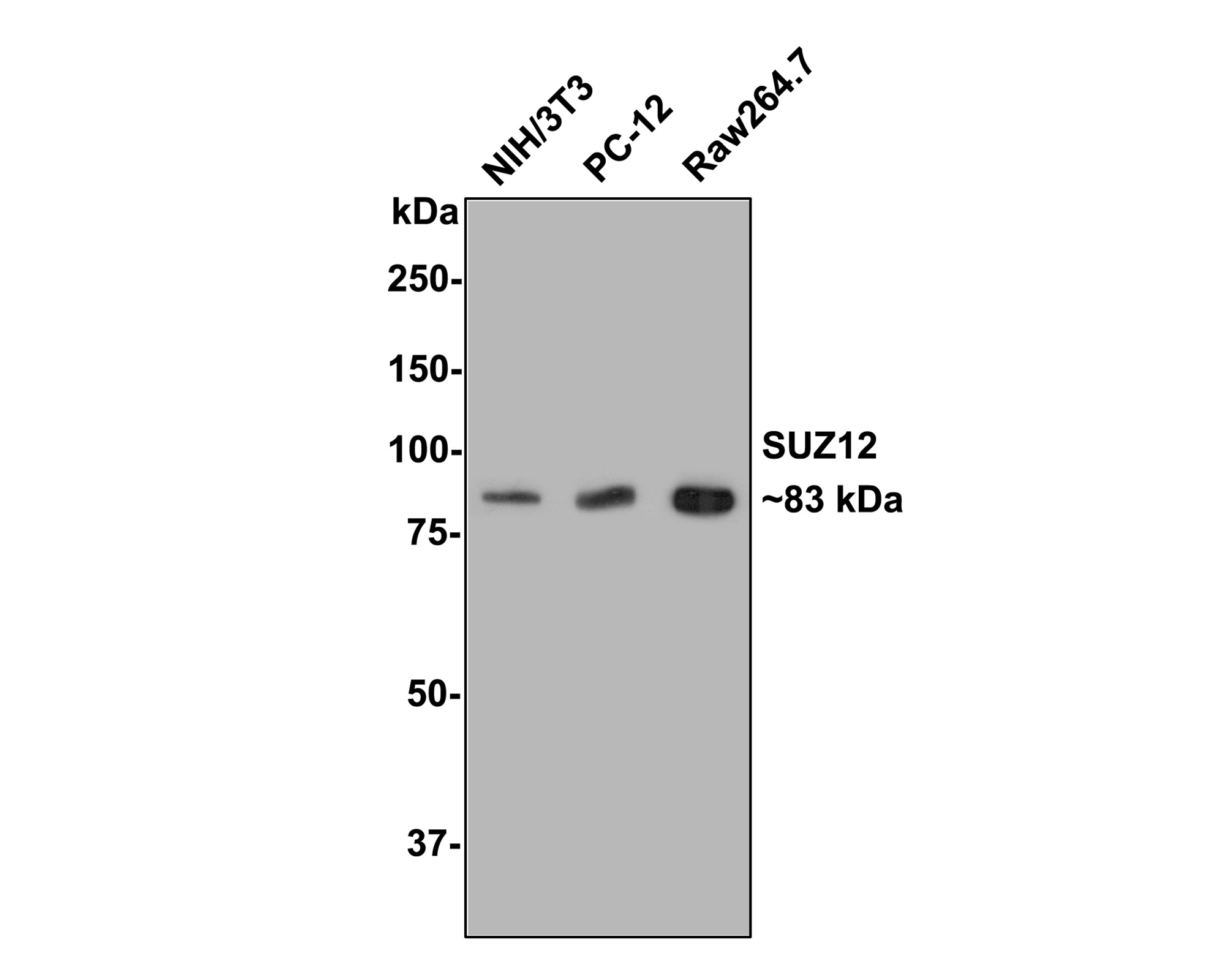
Fig2: Western blot analysis of SUZ12 on different lysates with Rabbit anti-SUZ12 antibody at 1/500 dilution. Lane 1: NIH/3T3 cell lysate, Lane 2: PC-12 cell lysate, Lane 3: Raw264.7 cell lysate, Lysates/proteins at 10 µg/Lane. Exposure time: 2 minutes; 8% SDS-PAGE gel. Proteins were transferred to a PVDF membrane and blocked with 5% NFDM/TBST for 1 hour at room temperature. The primary antibody at 1/500 dilution was used in 5% NFDM/TBST at room temperature for 2 hours. Goat Anti-Rabbit IgG - HRP Secondary Antibody at 1:300,000 dilution was used for 1 hour at room temperature.Application
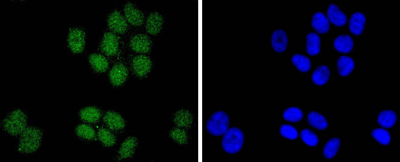
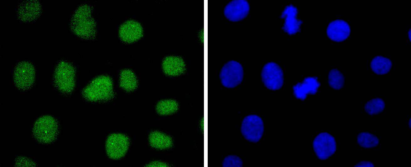
Fig3: ICC staining SUZ12 in MCF-7 cells (green). The nuclear counter stain is DAPI (blue). Cells were fixed in paraformaldehyde, permeabilised with 0.25% Triton X100/PBS.Application
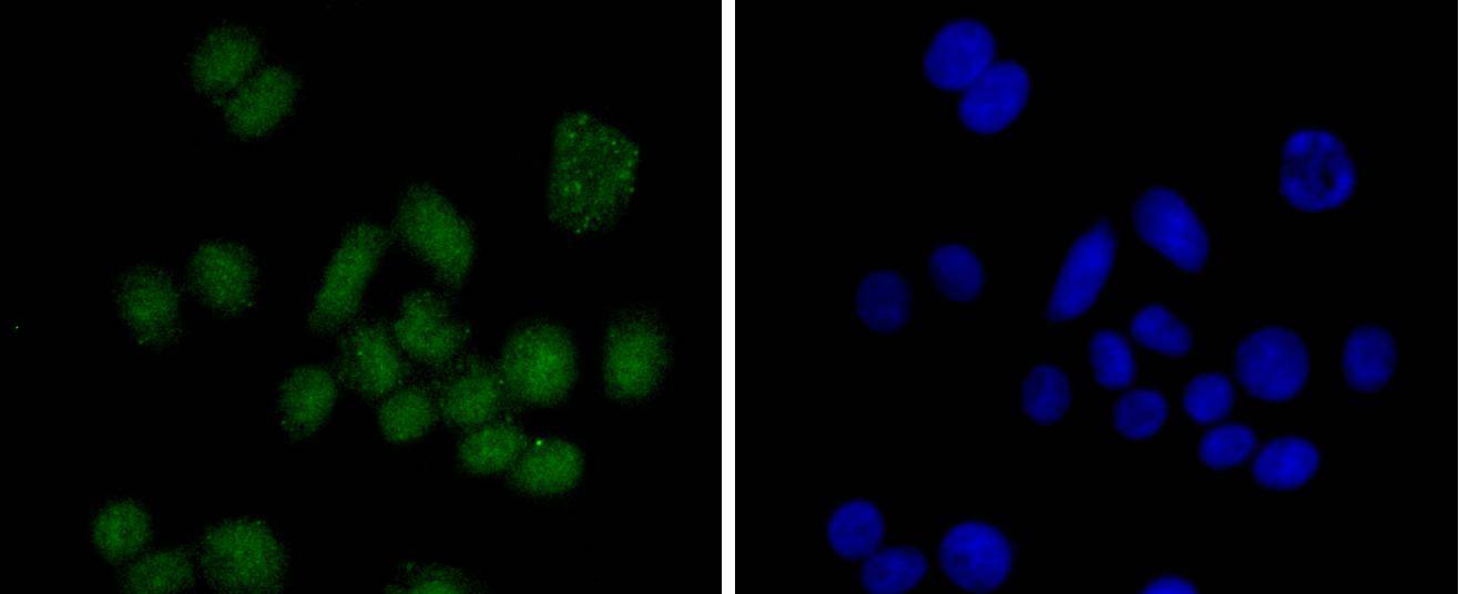
Fig4: ICC staining SUZ12 in SW480 cells (green). The nuclear counter stain is DAPI (blue). Cells were fixed in paraformaldehyde, permeabilised with 0.25% Triton X100/PBS.Application
Fig5: ICC staining SUZ12 in SKOV-3 cells (green). The nuclear counter stain is DAPI (blue). Cells were fixed in paraformaldehyde, permeabilised with 0.25% Triton X100/PBS.| Product Name | SUZ12 Recombinant Rabbit Monoclonal Antibody |
|---|---|
| Antibody Type | Primary Antibodies |
| Product description | The Polycomb group (PcG) genes contribute to the maintenance of cell identity, cell cycle regulation and oncogenesis. The mammalian PcG proteins are regulatory proteins important for Hox gene expression, axial skeleton development and the control of proliferation and survival of hematopoietic cells. By inducing changes in chromatin structure, the PcG proteins are part of a cellular memory system that is responsible for gene activity being inherited to progeny cells. PcG proteins silence gene expression through the formation of multimeric protein complexes with different compositions. Manipulating the expression-levels of various PcG proteins in mammalian cell lines results in cellular transformation, which may be a link between the chromatin-associated PcG proteins and cancer. Polycomb protein SUZ12, also designated ChET 9 protein or joined-to-JAZF1 protein, is a nuclear protein belonging to the VEFS (VRN2-EMF2-FIS2-SUZ12) family. SUZ12 has been detected at the breakpoints of a certain recurrent chromosomal translocation which has been reported in endometrial stromal sarcoma. It is a component of the PRC2 complex, composed of EED, EZH2, SUZ12/JJAZ1, RBBP4 and RBBP7. |
| Immunogen | Recombinant protein within human SUZ12 aa 1-150/739. |
| Clonality | monoclonal |
|---|---|
| Isotype | IgG |
| Host Species | Recombinant rabbit |
| Tested Applications | ICC/IFWB |
| WB:1:1,000-1:2,000 ICC:1:50-1:200 |
|
| Species Reactivity | HumanMouseRat |
| Concentration | 1mg/ml |
| Purification | Protein A |
| Alternative Names | ChET 9 protein antibody CHET9 antibody Chromatin precipitated E2F target 9 protein antibody JJAZ1 antibody Joined to JAZF1 protein antibody KIAA0160 antibody Polycomb protein SUZ12 antibody Suppressor of zeste 12 homolog antibody Suppressor of zeste 12 protein homolog antibody SUZ12 antibody SUZ12 polycomb repressive complex 2 subunit antibody SUZ12_HUMAN antibody |
|---|---|
| Molecular Weight(MW) | 83 kDa |
| Cellular Localization | Nucleus. |
Application
Fig1: Western blot analysis of SUZ12 on MCF-7 cells lysates using anti-SUZ12 antibody at 1/1,000 dilution.
Application
Fig2: Western blot analysis of SUZ12 on different lysates with Rabbit anti-SUZ12 antibody at 1/500 dilution. Lane 1: NIH/3T3 cell lysate, Lane 2: PC-12 cell lysate, Lane 3: Raw264.7 cell lysate, Lysates/proteins at 10 µg/Lane. Exposure time: 2 minutes; 8% SDS-PAGE gel. Proteins were transferred to a PVDF membrane and blocked with 5% NFDM/TBST for 1 hour at room temperature. The primary antibody at 1/500 dilution was used in 5% NFDM/TBST at room temperature for 2 hours. Goat Anti-Rabbit IgG - HRP Secondary Antibody at 1:300,000 dilution was used for 1 hour at room temperature.
Application
Fig3: ICC staining SUZ12 in MCF-7 cells (green). The nuclear counter stain is DAPI (blue). Cells were fixed in paraformaldehyde, permeabilised with 0.25% Triton X100/PBS.
Application
Fig4: ICC staining SUZ12 in SW480 cells (green). The nuclear counter stain is DAPI (blue). Cells were fixed in paraformaldehyde, permeabilised with 0.25% Triton X100/PBS.
Application
Fig5: ICC staining SUZ12 in SKOV-3 cells (green). The nuclear counter stain is DAPI (blue). Cells were fixed in paraformaldehyde, permeabilised with 0.25% Triton X100/PBS.| Positive Control | SW480, MCF-7, Hela, SKOV-3. |
|---|---|
| Application Notes | WB:1:1,000-1:2,000 ICC:1:50-1:200 |
| Form | Liquid |
|---|---|
| Storage Instructions | Store at +4℃ after thawing. Aliquot store at -20℃ or -80℃. Avoid repeated freeze / thaw cycles. |
| Storage Buffer | 1*TBS (pH7.4), 1%BSA, 40%Glycerol. Preservative: 0.05% Sodium Azide. |


