Application
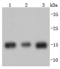
Fig1: Western blot analysis of Histone H3 on different lysates using anti-Histone H3 antibody at 1/1,000 dilution. Positive control: Lane 1: Hela Lane 2: MCF-7 Lane 3: NIH/3T3Application
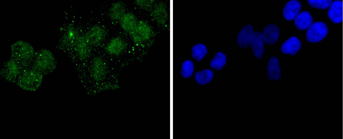
Fig2: ICC staining Histone H3 in Hela cells (green). The nuclear counter stain is DAPI (blue). Cells were fixed in paraformaldehyde, permeabilised with 0.25% Triton X100/PBS.Application
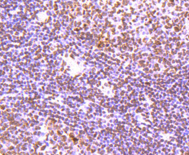
Fig3: Immunohistochemical analysis of paraffin-embedded human tonsil tissue using anti-Histone H3 antibody. Counter stained with hematoxylin.Application
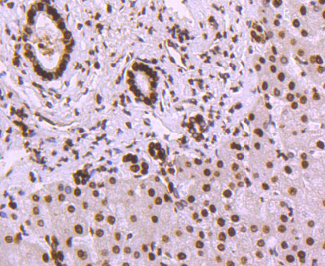
Fig4: Immunohistochemical analysis of paraffin-embedded human liver tissue using anti-Histone H3 antibody. Counter stained with hematoxylin.Application
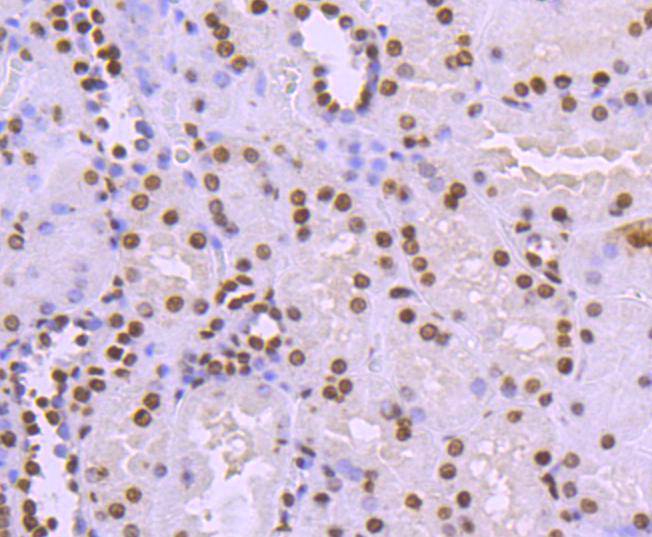
Fig5: Immunohistochemical analysis of paraffin-embedded human kidney tissue using anti-Histone H3 antibody. Counter stained with hematoxylin.Application
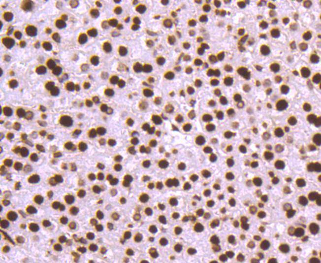
Fig6: Immunohistochemical analysis of paraffin-embedded mouse liver tissue using anti-Histone H3 antibody. Counter stained with hematoxylin.Application
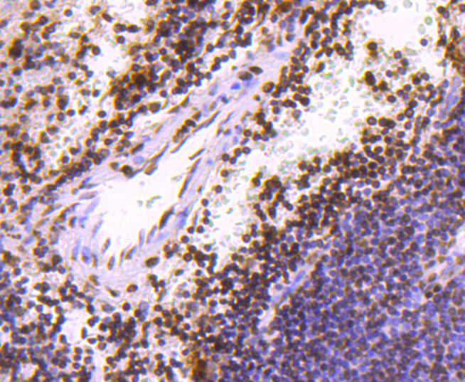
Fig7: Immunohistochemical analysis of paraffin-embedded human spleen tissue using anti-Histone H3 antibody. Counter stained with hematoxylin.Application
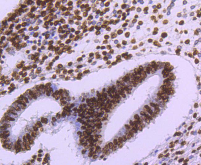
Fig8: Immunohistochemical analysis of paraffin-embedded human uterus tissue using anti-Histone H3 antibody. Counter stained with hematoxylin.| Product Name | Histone H3 [JJ090-07] |
|---|---|
| Antibody Type | Primary Antibodies |
| Antigen Alias | H3 histone family, member A antibody H3/A antibody H31_HUMAN antibody H3FA antibody Hist1h3a antibody HIST1H3B antibody HIST1H3C antibody HIST1H3D antibody HIST1H3E antibody HIST1H3F antibody HIST1H3G antibody HIST1H3H antibody HIST1H3I antibody HIST1H3J antibody histone 1, H3a antibody Histone cluster 1, H3a antibody Histone H3.1 antibody Histone H3/a antibody Histone H3/b antibody Histone H3/c antibody Histone H3/d antibody Histone H3/f antibody Histone H3/h antibody Histone H3/i antibody Histone H3/j antibody Histone H3/k antibody Histone H3/l antibody |
| Product description | In eukaryotes, DNA is wrapped around histone octamers to form the basic unit of chromatin structure. The octamer is composed of histones H2A, H2B, H3 and H4, and it associates with approximately 200 base pairs of DNA to form the nucleosome. The association of DNA with histones results in dense packing of chromatin, which restricts proteins involved in gene transcription from binding to DNA. p300 preferentially acetylates Histone H3 at lysines 14 and 18 and Histone H4 at lysines 5 and 8. PCAF in its native form, primarily acetylates Histone H3 at lysine 14 to a monoacetylated form, and less efficiently acetylates Histone H4 at lysine 8. Histone H4 may also be acetylated at lysines 12 and 16, and the involvement of acetylated H4 with Histones H2A, H2B and H3 suggests that acetylated histones may be involved in dynamic chromatin remodeling. |
| Immunogen | recombinant protein |
| Clonality | Monoclonal |
|---|---|
| Isotype | IgG |
| Host Species | Recombinant rabbit |
| Tested Applications | WBICC/IFIHCChIP |
| WB:1:1,000-1:2,000 ICC:1:50-1:200 IHC:1:100-1:500 |
|
| Species Reactivity | HumanMouseRat |
| Concentration | 1mg/ml |
| Alternative Names | H3 histone family member A antibody H3/A antibody H31_HUMAN antibody H3FA antibody Hist1h3a antibody HIST1H3B antibody HIST1H3C antibody HIST1H3D antibody HIST1H3E antibody HIST1H3F antibody HIST1H3G antibody HIST1H3H antibody HIST1H3I antibody HIST1H3J antibody histone 1 H3a antibody Histone cluster 1 H3a antibody Histone H3.1 antibody Histone H3/a antibody Histone H3/b antibody Histone H3/c antibody Histone H3/d antibody Histone H3/f antibody Histone H3/h antibody Histone H3/i antibody Histone H3/j antibody Histone H3/k antibody Histone H3/l antibody |
|---|---|
| Molecular Weight(MW) | 15 kDa |
| Cellular Localization | Nucleus, Chromosome. |
Application
Fig1: Western blot analysis of Histone H3 on different lysates using anti-Histone H3 antibody at 1/1,000 dilution. Positive control: Lane 1: Hela Lane 2: MCF-7 Lane 3: NIH/3T3
Application
Fig2: ICC staining Histone H3 in Hela cells (green). The nuclear counter stain is DAPI (blue). Cells were fixed in paraformaldehyde, permeabilised with 0.25% Triton X100/PBS.
Application
Fig3: Immunohistochemical analysis of paraffin-embedded human tonsil tissue using anti-Histone H3 antibody. Counter stained with hematoxylin.
Application
Fig4: Immunohistochemical analysis of paraffin-embedded human liver tissue using anti-Histone H3 antibody. Counter stained with hematoxylin.
Application
Fig5: Immunohistochemical analysis of paraffin-embedded human kidney tissue using anti-Histone H3 antibody. Counter stained with hematoxylin.
Application
Fig6: Immunohistochemical analysis of paraffin-embedded mouse liver tissue using anti-Histone H3 antibody. Counter stained with hematoxylin.
Application
Fig7: Immunohistochemical analysis of paraffin-embedded human spleen tissue using anti-Histone H3 antibody. Counter stained with hematoxylin.
Application
Fig8: Immunohistochemical analysis of paraffin-embedded human uterus tissue using anti-Histone H3 antibody. Counter stained with hematoxylin.| Positive Control | NIH/3T3, MCF-7, Hela, human tonsil tissue, human liver tissue, human kidney tissue, human uterus tissue, mouse liver tissue. |
|---|---|
| Application Notes | WB:1:1,000-1:2,000 ICC:1:50-1:200 IHC:1:100-1:500 |
| Form | Liquid |
|---|---|
| Storage Instructions | Store at +4℃ after thawing. Aliquot store at -20℃ or -80℃. Avoid repeated freeze / thaw cycles. |
| Storage Buffer | 1*TBS (pH7.4), 1%BSA, 40%Glycerol. Preservative: 0.05% Sodium Azide. |


