WB
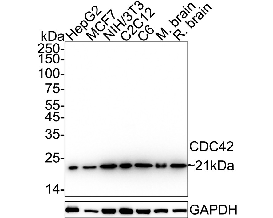
Western blot analysis of CDC42 on different lysates with Rabbit anti-CDC42 antibody at 1/1,000 dilution. Lane 1: HepG2 cell lysate, Lane 2: MCF7 cell lysate, Lane 3: NIH/3T3 cell lysate, Lane 4: C2C12 cell lysate, Lane 5: C6 cell lysate, Lane 6: Mouse brain tissue lysate, Lane 7: Rat brain tissue lysate, Lysates/proteins at 20 µg/Lane. Exposure time: 8 seconds; 4-20% SDS-PAGE gel. Proteins were transferred to a PVDF membrane and blocked with 5% NFDM/TBST for 1 hour at room temperature. The primary antibody at 1/1,000 dilution was used in 5% NFDM/TBST at 4℃ overnight. Goat Anti-Rabbit IgG - HRP Secondary Antibody at 1/50,000 dilution was used for 1 hour at room temperature.ICC/IF
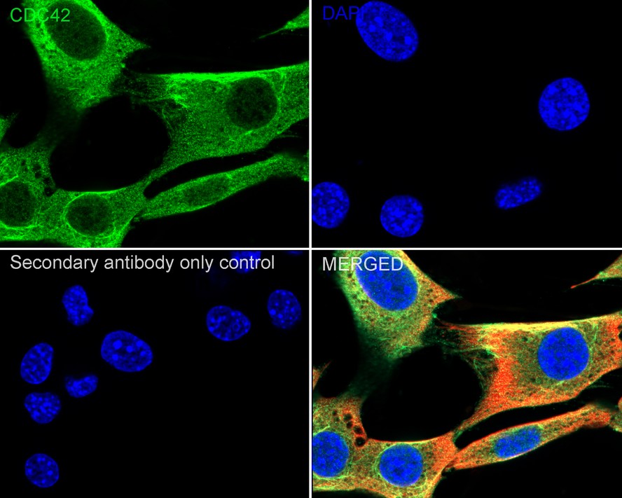
Immunocytochemistry analysis of NIH/3T3 cells labeling CDC42 with Rabbit anti-CDC42 antibody at 1/500 dilution. Cells were fixed in 4% paraformaldehyde for 15 minutes at room temperature, permeabilized with 0.1% Triton X-100 in PBS for 15 minutes at room temperature, then blocked with 1% BSA in 10% negative goat serum for 1 hour at room temperature. Cells were then incubated with Rabbit anti-CDC42 antibody at 1/500 dilution in 1% BSA in PBST overnight at 4 ℃. Goat Anti-Rabbit IgG H&L (488) was used as the secondary antibody at 1/1,000 dilution. PBS instead of the primary antibody was used as the secondary antibody only control. Nuclear DNA was labelled in blue with DAPI. Beta tubulin (red) was stained at 1/100 dilution overnight at +4℃. Goat Anti-Mouse IgG H&L (594) was used as the secondary antibody at 1/1,000 dilution.ICC/IF
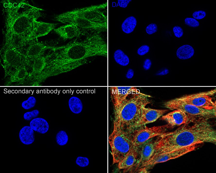
Immunocytochemistry analysis of C6 cells labeling CDC42 with Rabbit anti-CDC42 antibody at 1/500 dilution. Cells were fixed in 4% paraformaldehyde for 15 minutes at room temperature, permeabilized with 0.1% Triton X-100 in PBS for 15 minutes at room temperature, then blocked with 1% BSA in 10% negative goat serum for 1 hour at room temperature. Cells were then incubated with Rabbit anti-CDC42 antibody at 1/500 dilution in 1% BSA in PBST overnight at 4 ℃. Goat Anti-Rabbit IgG H&L (488) was used as the secondary antibody at 1/1,000 dilution. PBS instead of the primary antibody was used as the secondary antibody only control. Nuclear DNA was labelled in blue with DAPI. Beta tubulin ( red) was stained at 1/100 dilution overnight at +4℃. Goat Anti-Mouse IgG H&L (594) was used as the secondary antibody at 1/1,000 dilution.FC
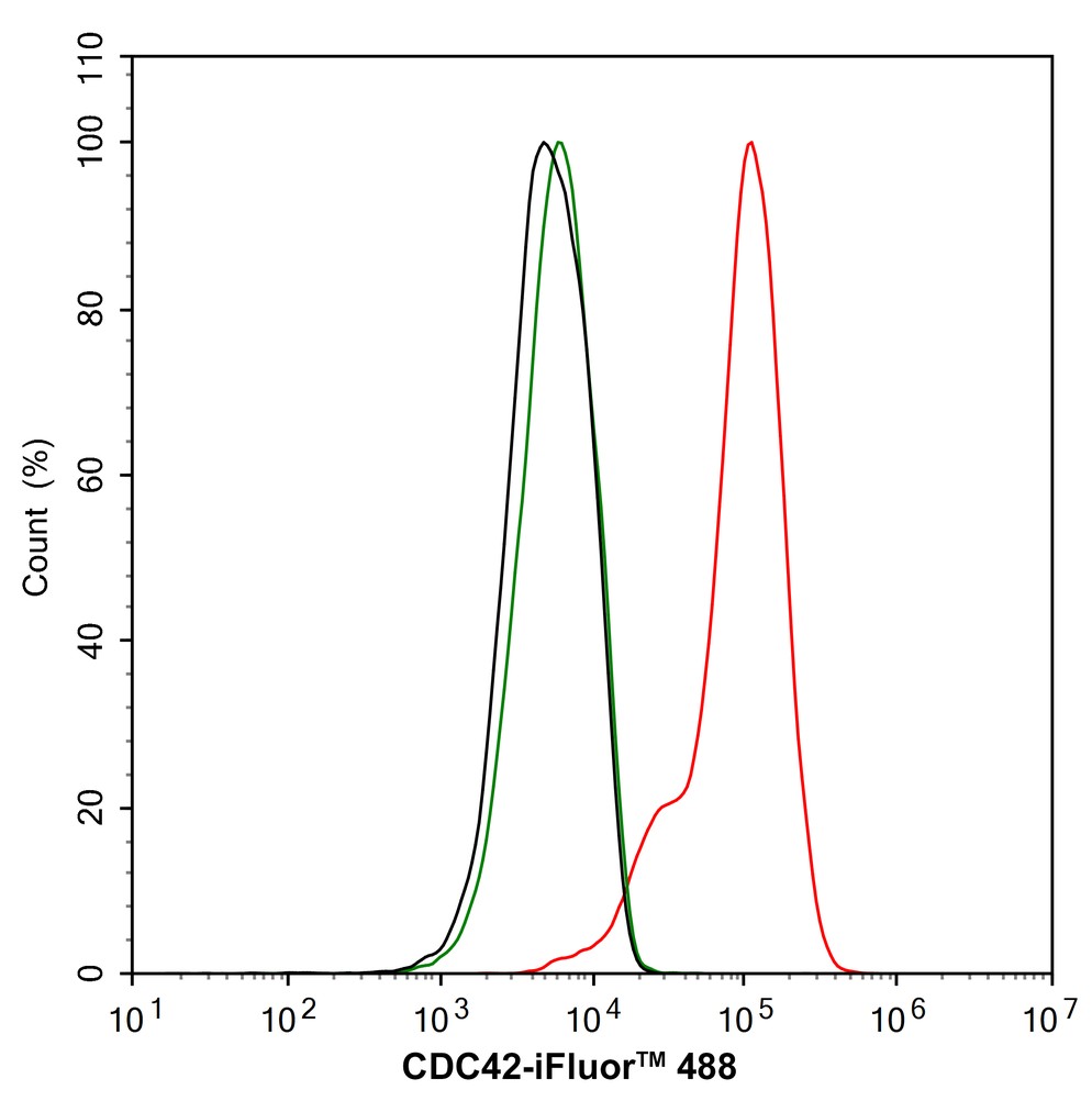
Flow cytometric analysis of HepG2 cells labeling CDC42. Cells were fixed and permeabilized. Then stained with the primary antibody (1/1,000) (red) compared with Rabbit IgG Isotype Control (green). After incubation of the primary antibody at +4℃ for an hour, the cells were stained with a iFluor™ 488 conjugate-Goat anti-Rabbit IgG Secondary antibody at 1/1,000 dilution for 30 minutes at +4℃. Unlabelled sample was used as a control (cells without incubation with primary antibody; black).| Product Name | CDC42 Recombinant Rabbit Monoclonal Antibody |
|---|---|
| Antibody Type | Primary Antibodies |
| Product description | The superfamily of GTP-binding proteins, for which the Ras proteins are prototypes, has been implicated in regulation of diverse biological activities involving various aspects of cell growth and division. One mammalian member of the family, Cdc42, has an amino acid sequence that is similar to those of various members of the Ras superfamily proteins, including N-, K- and H-Ras, Rho proteins and the Rac proteins. On the basis of in vitro phosphorylation studies, it has been suggested that human Cdc42 may function in the signaling pathway of the EGF receptor or related growth factor receptor protein kinases. The Dbl oncogene has been shown to specifically catalyze dissociation of GDP from human Cdc42. |
| Immunogen | Recombinant protein within Human CDC42 aa 30-191 / 191. |
| Clonality | Monoclonal |
|---|---|
| Isotype | IgG |
| Host Species | Recombinant rabbit |
| Tested Applications | FCICC/IFWB |
| WB:1:1000 ICC/IF:1:500 FC:1:1000 |
|
| Species Reactivity | HumanMouseRat |
| Concentration | 1mg/ml |
| Purification | Protein A |
| Alternative Names | CDC42 antibody
CDC42_HUMAN antibody
CDC42Hs antibody
Cell division control protein 42 homolog antibody
Cell division cycle 42 (GTP binding protein 25kDa) antibody
Cell division cycle 42 antibody
dJ224A6.1.1 (cell division cycle 42 (GTP-binding protein 25kD)) antibody dJ224A6.1.2 (cell division cycle 42 (GTP-binding protein 25kD)) antibody G25K antibody G25K GTP-binding protein antibody Growth regulating protein antibody GTP binding protein 25kDa antibody Small GTP binding protein CDC42 antibody TKS antibody |
|---|---|
| Molecular Weight(MW) | 21kDa |
| Cellular Localization | Cell membrane, Cell projection, Cytoplasm, Cytoskeleton, Membrane. |
WB
Western blot analysis of CDC42 on different lysates with Rabbit anti-CDC42 antibody at 1/1,000 dilution. Lane 1: HepG2 cell lysate, Lane 2: MCF7 cell lysate, Lane 3: NIH/3T3 cell lysate, Lane 4: C2C12 cell lysate, Lane 5: C6 cell lysate, Lane 6: Mouse brain tissue lysate, Lane 7: Rat brain tissue lysate, Lysates/proteins at 20 µg/Lane. Exposure time: 8 seconds; 4-20% SDS-PAGE gel. Proteins were transferred to a PVDF membrane and blocked with 5% NFDM/TBST for 1 hour at room temperature. The primary antibody at 1/1,000 dilution was used in 5% NFDM/TBST at 4℃ overnight. Goat Anti-Rabbit IgG - HRP Secondary Antibody at 1/50,000 dilution was used for 1 hour at room temperature.
ICC/IF
Immunocytochemistry analysis of NIH/3T3 cells labeling CDC42 with Rabbit anti-CDC42 antibody at 1/500 dilution. Cells were fixed in 4% paraformaldehyde for 15 minutes at room temperature, permeabilized with 0.1% Triton X-100 in PBS for 15 minutes at room temperature, then blocked with 1% BSA in 10% negative goat serum for 1 hour at room temperature. Cells were then incubated with Rabbit anti-CDC42 antibody at 1/500 dilution in 1% BSA in PBST overnight at 4 ℃. Goat Anti-Rabbit IgG H&L (488) was used as the secondary antibody at 1/1,000 dilution. PBS instead of the primary antibody was used as the secondary antibody only control. Nuclear DNA was labelled in blue with DAPI. Beta tubulin (red) was stained at 1/100 dilution overnight at +4℃. Goat Anti-Mouse IgG H&L (594) was used as the secondary antibody at 1/1,000 dilution.
ICC/IF
Immunocytochemistry analysis of C6 cells labeling CDC42 with Rabbit anti-CDC42 antibody at 1/500 dilution. Cells were fixed in 4% paraformaldehyde for 15 minutes at room temperature, permeabilized with 0.1% Triton X-100 in PBS for 15 minutes at room temperature, then blocked with 1% BSA in 10% negative goat serum for 1 hour at room temperature. Cells were then incubated with Rabbit anti-CDC42 antibody at 1/500 dilution in 1% BSA in PBST overnight at 4 ℃. Goat Anti-Rabbit IgG H&L (488) was used as the secondary antibody at 1/1,000 dilution. PBS instead of the primary antibody was used as the secondary antibody only control. Nuclear DNA was labelled in blue with DAPI. Beta tubulin ( red) was stained at 1/100 dilution overnight at +4℃. Goat Anti-Mouse IgG H&L (594) was used as the secondary antibody at 1/1,000 dilution.
FC
Flow cytometric analysis of HepG2 cells labeling CDC42. Cells were fixed and permeabilized. Then stained with the primary antibody (1/1,000) (red) compared with Rabbit IgG Isotype Control (green). After incubation of the primary antibody at +4℃ for an hour, the cells were stained with a iFluor™ 488 conjugate-Goat anti-Rabbit IgG Secondary antibody at 1/1,000 dilution for 30 minutes at +4℃. Unlabelled sample was used as a control (cells without incubation with primary antibody; black).| Application Notes | WB:1:1000 ICC/IF:1:500 FC:1:1000 |
|---|
| Form | Liquid |
|---|---|
| Storage Instructions | Store at +4℃ after thawing. Aliquot store at -20℃ or -80℃. Avoid repeated freeze / thaw cycles. |
| Storage Buffer | 1*TBS (pH7.4), 1%BSA, 40%Glycerol. Preservative: 0.05% Sodium Azide. |


