Application
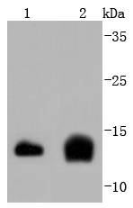
Fig1: Western blot analysis of Cystatin C on different lysates using anti-Cystatin C antibody at 1/1,000 dilution. Positive control: Lane 1: Hela Lane 2: Mouse spleenApplication
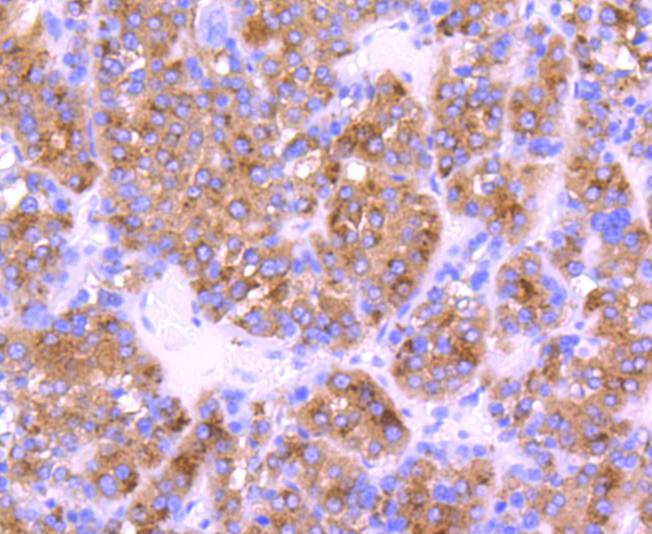
Fig2: Immunohistochemical analysis of paraffin-embedded human liver cancer tissue using anti-Cystatin C antibody. Counter stained with hematoxylin.Application
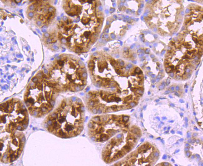
Fig3: Immunohistochemical analysis of paraffin-embedded human kidney tissue using anti-Cystatin C antibody. Counter stained with hematoxylin.Application
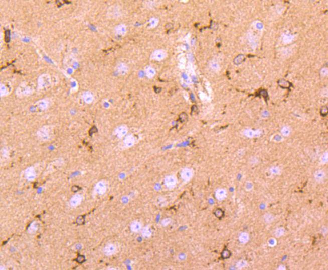
Fig4: Immunohistochemical analysis of paraffin-embedded mouse brain tissue using anti-Cystatin C antibody. Counter stained with hematoxylin.Application
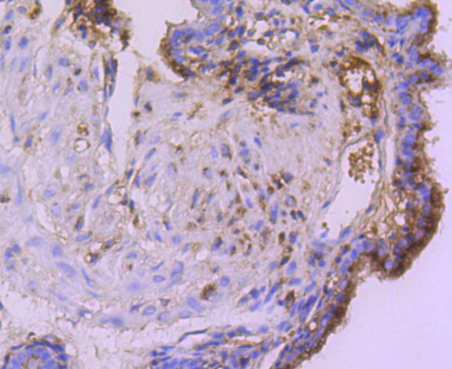
Fig5: Immunohistochemical analysis of paraffin-embedded mouse placenta tissue using anti-Cystatin C antibody. Counter stained with hematoxylin.Application
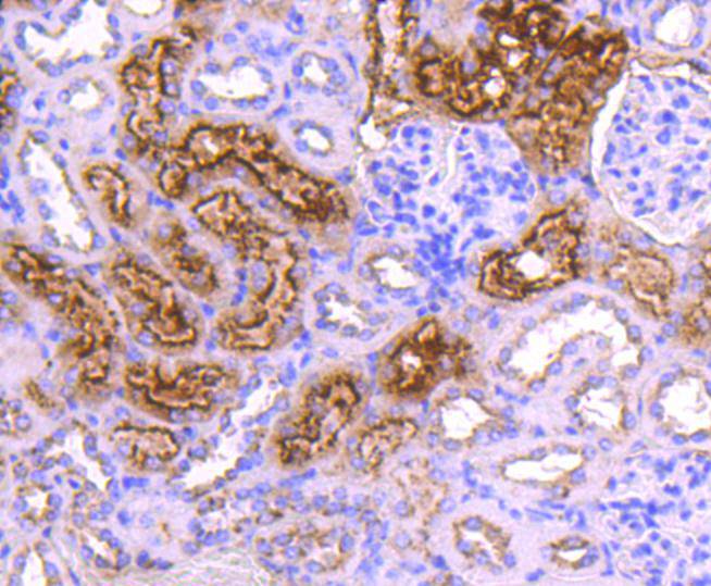
Fig6: Immunohistochemical analysis of paraffin-embedded mouse kidney tissue using anti-Cystatin C antibody. Counter stained with hematoxylin.| Product Name | Cystatin C [JJ09-16] |
|---|---|
| Antibody Type | Primary Antibodies |
| Antigen Alias | AD 8 antibody AD8 antibody Amyloid angiopathy and cerebral hemorrhage antibody ARMD11 antibody bA218C14.4 (cystatin C) antibody bA218C14.4 antibody Cst 3 antibody Cst3 antibody CST3 protein antibody Cystatin 3 antibody Cystatin-3 antibody Cystatin-C antibody Cystatin3 antibody CystatinC antibody CYTC_HUMAN antibody Epididymis secretory protein Li 2 antibody Gamma trace antibody Gamma-trace antibody HCCAA antibody HEL S 2 antibody MGC117328 antibody Neuroendocrine basic polypeptide antibody Post gamma globulin antibody Post-gamma-globulin antibody |
| Product description | Cystatin C is a cysteine (thiol) protease inhibitor that belongs to the type II cystatin gene superfamily and is the most abundant extracellular inhibitor of cysteine proteases. Cystatin C is a constitutively secreted, amyloidogenic protein, which forms a two-fold symmetric dimer and modulates both cysteine protease activity and the expression of class II MHC molecules. Expression of cystatin C is an indicator of kidney function and glomerular filtration rate. Mutations in the cystatin C gene can lead to protein aggregates, which are implicated in hereditary amyloid angiopathy (HCCAA) and cerebral hemorrhage. Although both wild-type and mutant cystatin C are capable of forming concentration dependent inactive dimers, mutant cystatin C dimerizes at lower concentrations and is more susceptible to serine proteases, which may facilitate aggregation. In neuronal cells, oxidative stress stimulates expression of cystatin C, which may positively regulate apoptosis. |
| Immunogen | recombinant protein |
| Clonality | Monoclonal |
|---|---|
| Isotype | IgG |
| Host Species | Recombinant rabbit |
| Tested Applications | WBIHC |
| WB:1:1,000-1:2,000 IHC:1:50-1:200 |
|
| Species Reactivity | HumanMouseRat |
| Concentration | 1mg/ml |
| Alternative Names | AD 8 antibody AD8 antibody Amyloid angiopathy and cerebral hemorrhage antibody ARMD11 antibody bA218C14.4 (cystatin C) antibody bA218C14.4 antibody Cst 3 antibody Cst3 antibody CST3 protein antibody Cystatin 3 antibody Cystatin-3 antibody Cystatin-C antibody Cystatin3 antibody CystatinC antibody CYTC_HUMAN antibody Epididymis secretory protein Li 2 antibody Gamma trace antibody Gamma-trace antibody HCCAA antibody HEL S 2 antibody MGC117328 antibody Neuroendocrine basic polypeptide antibody Post gamma globulin antibody Post-gamma-globulin antibody |
|---|---|
| Molecular Weight(MW) | 16 kDa |
| Cellular Localization | Secreted. |
Application
Fig1: Western blot analysis of Cystatin C on different lysates using anti-Cystatin C antibody at 1/1,000 dilution. Positive control: Lane 1: Hela Lane 2: Mouse spleen
Application
Fig2: Immunohistochemical analysis of paraffin-embedded human liver cancer tissue using anti-Cystatin C antibody. Counter stained with hematoxylin.
Application
Fig3: Immunohistochemical analysis of paraffin-embedded human kidney tissue using anti-Cystatin C antibody. Counter stained with hematoxylin.
Application
Fig4: Immunohistochemical analysis of paraffin-embedded mouse brain tissue using anti-Cystatin C antibody. Counter stained with hematoxylin.
Application
Fig5: Immunohistochemical analysis of paraffin-embedded mouse placenta tissue using anti-Cystatin C antibody. Counter stained with hematoxylin.
Application
Fig6: Immunohistochemical analysis of paraffin-embedded mouse kidney tissue using anti-Cystatin C antibody. Counter stained with hematoxylin.| Positive Control | Hela, human liver cancer tissue, human kidney tissue, mouse brain tissue, mouse spleen tissue, mouse placenta tissue, mouse kidney tissue. |
|---|---|
| Application Notes | WB:1:1,000-1:2,000 IHC:1:50-1:200 |
| Form | Liquid |
|---|---|
| Storage Instructions | Store at +4℃ after thawing. Aliquot store at -20℃ or -80℃. Avoid repeated freeze / thaw cycles. |
| Storage Buffer | 1*TBS (pH7.4), 1%BSA, 40%Glycerol. Preservative: 0.05% Sodium Azide. |


