Application
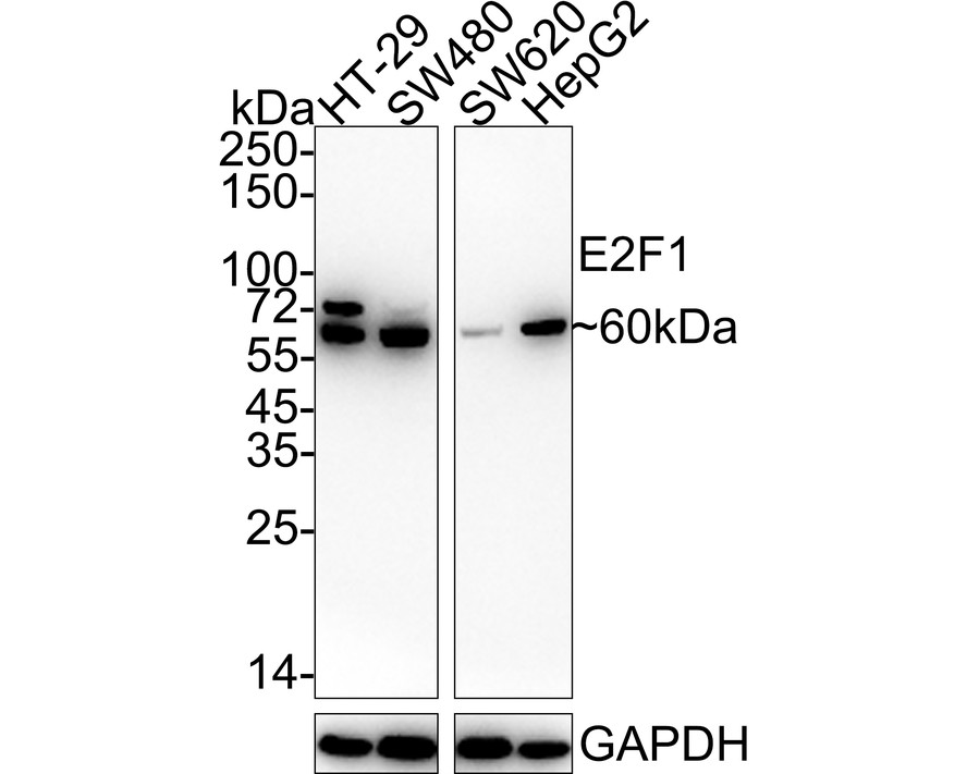
Fig1: Western blot analysis of E2F1 on different lysates with Rabbit anti-E2F1 antibody at 1/500 dilution. Lane 1: HT-29 cell lysate, Lane 2: SW480 cell lysate, Lane 3: SW620 cell lysate, Lane 4: HepG2 cell lysate, Lysates/proteins at 20 µg/Lane. Exposure time: 3 minutes; 4-20% SDS-PAGE gel. Proteins were transferred to a PVDF membrane and blocked with 5% NFDM/TBST for 1 hour at room temperature. The primary antibody at 1/500 dilution was used in 5% NFDM/TBST at 4℃ overnight. Goat Anti-Rabbit IgG - HRP Secondary Antibody at 1/50,000 dilution was used for 1 hour at room temperature.Application
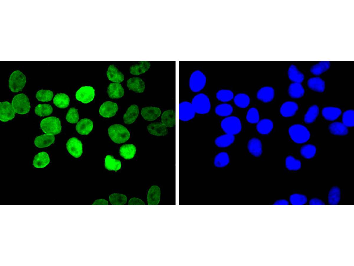
Fig2: ICC staining E2F1 in Hela cells (green). The nuclear counter stain is DAPI (blue). Cells were fixed in paraformaldehyde, permeabilised with 0.25% Triton X100/PBS.Application
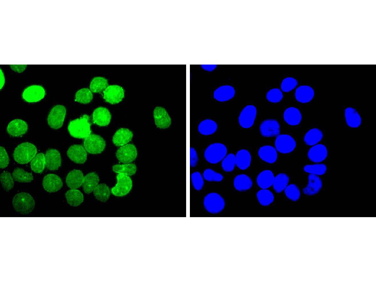
Fig3: ICC staining E2F1 in MCF-7 cells (green). The nuclear counter stain is DAPI (blue). Cells were fixed in paraformaldehyde, permeabilised with 0.25% Triton X100/PBS.Application
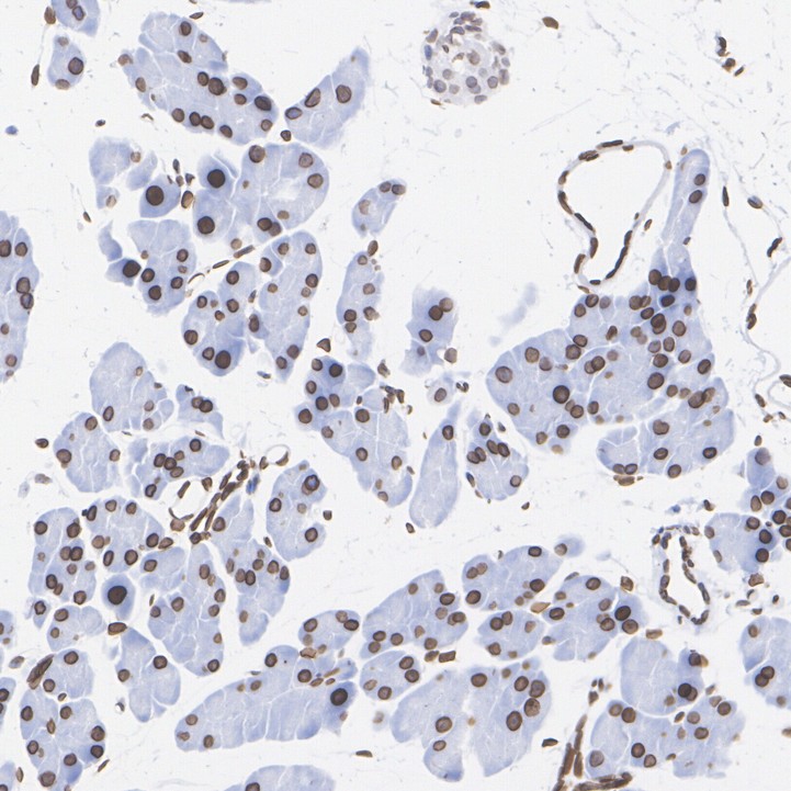
Fig4: Immunohistochemical analysis of paraffin-embedded mouse pancreas tissue with Rabbit anti-E2F1 antibody at 1/5,000 dilution. The section was pre-treated using heat mediated antigen retrieval with sodium citrate buffer (pH 6.0) for 2 minutes. The tissues were blocked in 1% BSA for 20 minutes at room temperature, washed with ddH2O and PBS, and then probed with the primary antibody at 1/5,000 dilution for 1 hour at room temperature. The detection was performed using an HRP conjugated compact polymer system. DAB was used as the chromogen. Tissues were counterstained with hematoxylin and mounted with DPX.Application
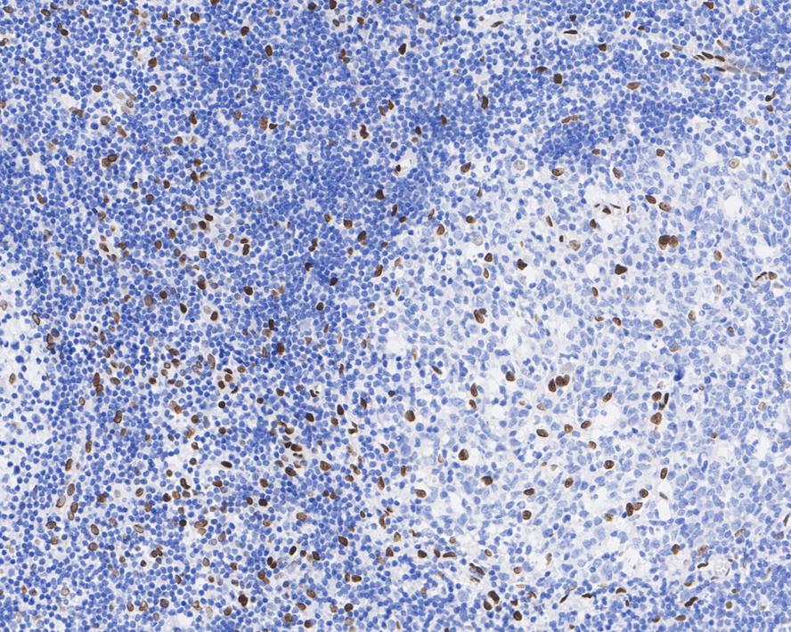
Fig5: Immunohistochemical analysis of paraffin-embedded human tonsil tissue with Rabbit anti-E2F1 antibody at 1/1,000 dilution. The section was pre-treated using heat mediated antigen retrieval with sodium citrate buffer (pH 6.0) for 2 minutes. The tissues were blocked in 1% BSA for 20 minutes at room temperature, washed with ddH2O and PBS, and then probed with the primary antibody at 1/1,000 dilution for 1 hour at room temperature. The detection was performed using an HRP conjugated compact polymer system. DAB was used as the chromogen. Tissues were counterstained with hematoxylin and mounted with DPX.Application
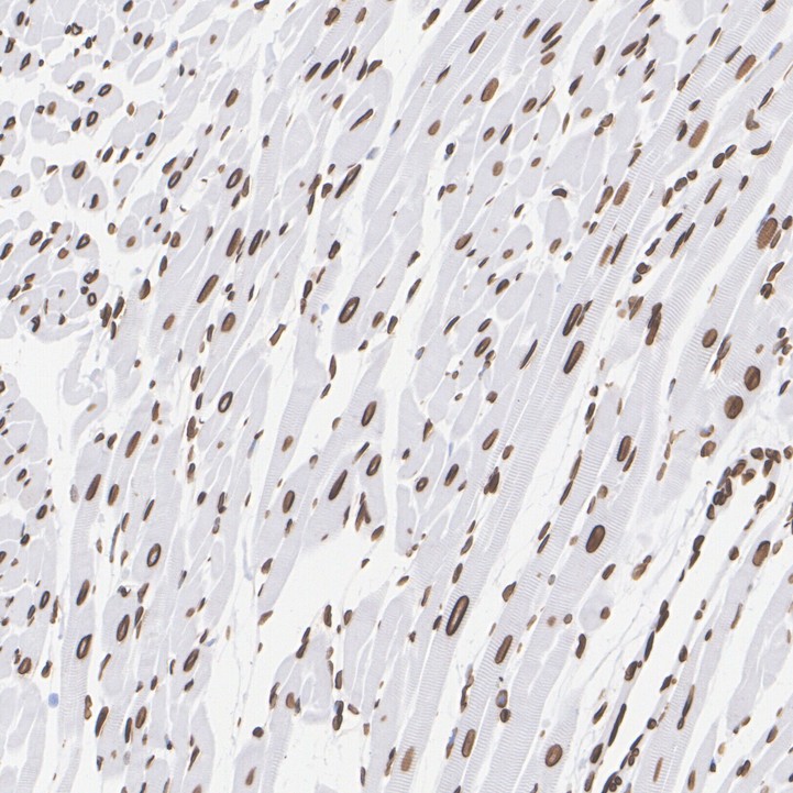
Fig6: Immunohistochemical analysis of paraffin-embedded rat heart tissue with Rabbit anti-E2F1 antibody at 1/5,000 dilution. The section was pre-treated using heat mediated antigen retrieval with sodium citrate buffer (pH 6.0) for 2 minutes. The tissues were blocked in 1% BSA for 20 minutes at room temperature, washed with ddH2O and PBS, and then probed with the primary antibody at 1/5,000 dilution for 1 hour at room temperature. The detection was performed using an HRP conjugated compact polymer system. DAB was used as the chromogen. Tissues were counterstained with hematoxylin and mounted with DPX.Application
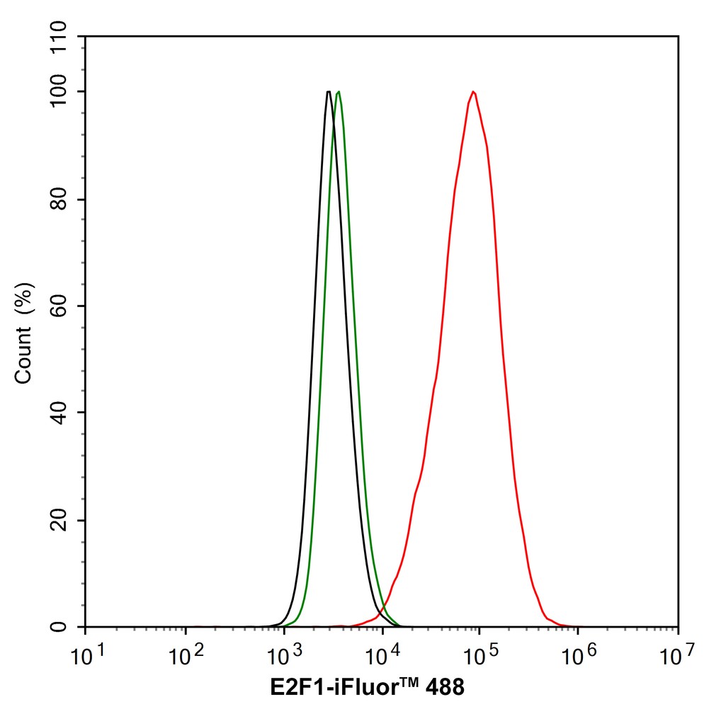
Fig7: Flow cytometric analysis of HepG2 cells labeling E2F1. Cells were fixed and permeabilized. Then stained with the primary antibody (1/1,000) (red) compared with Rabbit IgG Isotype Control (green). After incubation of the primary antibody at +4℃ for an hour, the cells were stained with a iFluor™ 488 conjugate-Goat anti-Rabbit IgG Secondary antibody at 1/1,000 dilution for 30 minutes at +4℃. Unlabelled sample was used as a control (cells without incubation with primary antibody; black).Application
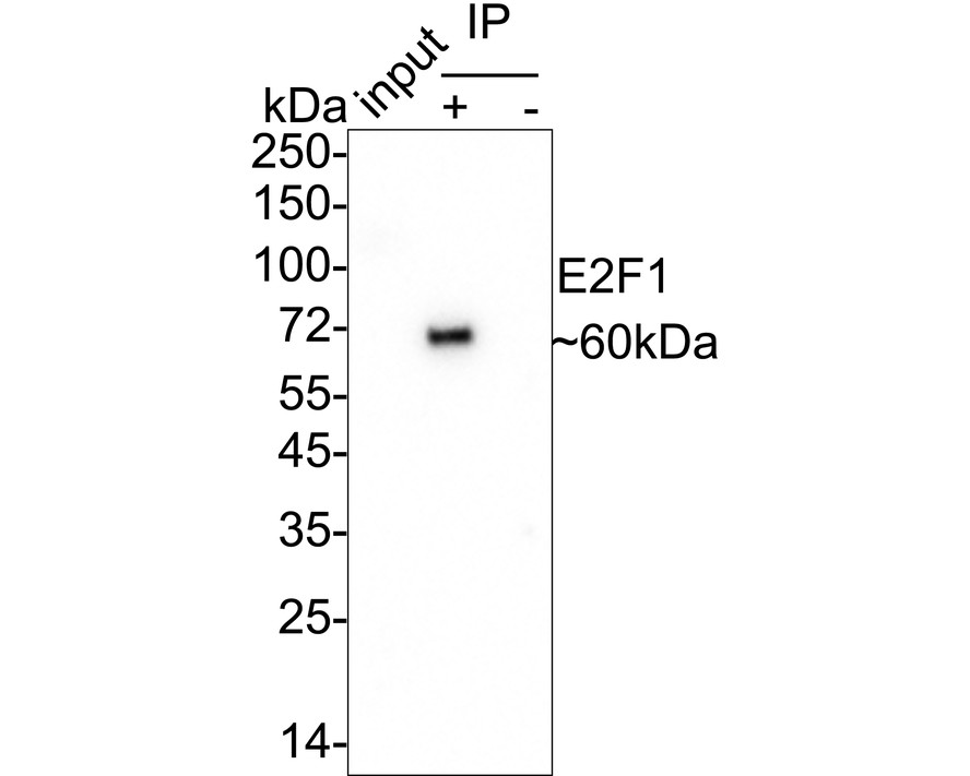
Fig8: E2F1 was immunoprecipitated from 0.2 mg HepG2 cell lysate with Rabbit anti-E2F1 antibody at 2 µg/25 µl agarose. Western blot was performed from the immunoprecipitate using Rabbit anti-E2F1 antibody at 1/1,000 dilution. Anti-Rabbit IgG for IP Nano-secondary antibody at 1/5,000 dilution was used for 1 hour at room temperature. Lane 1: HepG2 cell lysate (input), Lane 2:Rabbit anti-E2F1 antibody IP in HepG2 cell lysate, Lane 3: Rabbit IgG instead of Rabbit anti-E2F1 antibody in HepG2 cell lysate. Blocking/Dilution buffer: 5% NFDM/TBST. Exposure time: 26 seconds.| Product Name | E2F1 Recombinant Rabbit Monoclonal Antibody |
|---|---|
| Antibody Type | Primary Antibodies |
| Product description | The human retinoblastoma gene product appears to play an important role in the negative regulation of cell proliferation. Functional inactivation of Rb can be mediated either through mutation or as a consequence of interaction with DNA tumor virus-encoded proteins. Of all the Rb associations described to date, the identification of a complex between Rb and the transcription factor E2F most directly implicates Rb in regulation of cell proliferation. E2F was originally identified through its role in transcriptional activation of the adenovirus E2 promoter. Sequences homologous to the E2F binding site have been found upstream of a number of genes that encode proteins with putative functions in the G1 and S phases of the cell cycle. E2F-1 is a member of a broader family of transcription regulators including E2F-2, E2F-3, E2F-4, E2F-5, E2F-6 and E2F-7 each of which forms heterodimers with a second protein, DP-1, forming an "active" E2F transcriptional regulatory complex. |
| Immunogen | Synthetic peptide within Human E2F1 aa 371-404 / 437. |
| Clonality | monoclonal |
|---|---|
| Isotype | IgG |
| Host Species | Recombinant rabbit |
| Tested Applications | FCICC/IFIHCIPWB |
| WB:1:500-1:10000 ICC:1:100-1:500 IHC:1:1000-1:5000 FC:1:1000 IP:1-2μg/sample |
|
| Species Reactivity | HumanMouseRat |
| Concentration | 1mg/ml |
| Purification | Protein A |
| Gene Symbol | E2F1 |
|---|---|
| Gene Synonyms | RBP3 E2F-1 RBAP1 RBBP3 |
| Gene Full Name | E2F transcription factor 1 |
| Gene Summary | The protein encoded by this gene is a member of the E2F family of transcription factors. The E2F family plays a crucial role in the control of cell cycle and action of tumor suppressor proteins and is also a target of the transforming proteins of small DNA tumor viruses. The E2F proteins contain several evolutionally conserved domains found in most members of the family. These domains include a DNA binding domain, a dimerization domain which determines interaction with the differentiation regulated transcription factor proteins (DP), a transactivation domain enriched in acidic amino acids, and a tumor suppressor protein association domain which is embedded within the transactivation domain. This protein and another 2 members, E2F2 and E2F3, have an additional cyclin binding domain. This protein binds preferentially to retinoblastoma protein pRB in a cell-cycle dependent manner. It can mediate both cell proliferation and p53-dependent/independent apoptosis. [provided by RefSeq, Jul 2008] |
| Alternative Names | DmelCG6376 antibody Dmel_CG6376 antibody drosE2F1 antibody E(Sev-CycE)3A antibody E(var)3-93E antibody E2-promoter binding facto antibody E2F 1 antibody E2F transcription factor 1 antibody E2F-1 antibody E2f-PA antibody E2f-PB antibody E2f-PC antibody E2F1 antibody E2f1 E2F transcription factor 1 antibody E2F1_HUMAN antibody Evar(3)164 antibody KIAA4009 antibody l(3)07172 antibody l(3)j3B1 antibody l(3)j3C2 antibody l(3)rM729 antibody mKIAA4009 antibody OTTHUMP00000030661 antibody PBR3 antibody PRB binding protein E2F 1 antibody PRB-binding protein E2F-1 antibody RBAP 1 antibody RBAP-1 antibody RBAP1 antibody RBBP-3 antibody RBBP3 antibody RBP 3 antibody RBP3 antibody Retinoblastoma-associated protein 1 antibody Retinoblastoma-binding protein 3 antibody Transcription factor E2F1 antibody |
| Molecular Weight(MW) | 47kDa(Observed band size: 60kDa) |
| Cellular Localization | Nucleus. |
Application
Fig1: Western blot analysis of E2F1 on different lysates with Rabbit anti-E2F1 antibody at 1/500 dilution. Lane 1: HT-29 cell lysate, Lane 2: SW480 cell lysate, Lane 3: SW620 cell lysate, Lane 4: HepG2 cell lysate, Lysates/proteins at 20 µg/Lane. Exposure time: 3 minutes; 4-20% SDS-PAGE gel. Proteins were transferred to a PVDF membrane and blocked with 5% NFDM/TBST for 1 hour at room temperature. The primary antibody at 1/500 dilution was used in 5% NFDM/TBST at 4℃ overnight. Goat Anti-Rabbit IgG - HRP Secondary Antibody at 1/50,000 dilution was used for 1 hour at room temperature.
Application
Fig2: ICC staining E2F1 in Hela cells (green). The nuclear counter stain is DAPI (blue). Cells were fixed in paraformaldehyde, permeabilised with 0.25% Triton X100/PBS.
Application
Fig3: ICC staining E2F1 in MCF-7 cells (green). The nuclear counter stain is DAPI (blue). Cells were fixed in paraformaldehyde, permeabilised with 0.25% Triton X100/PBS.
Application
Fig4: Immunohistochemical analysis of paraffin-embedded mouse pancreas tissue with Rabbit anti-E2F1 antibody at 1/5,000 dilution. The section was pre-treated using heat mediated antigen retrieval with sodium citrate buffer (pH 6.0) for 2 minutes. The tissues were blocked in 1% BSA for 20 minutes at room temperature, washed with ddH2O and PBS, and then probed with the primary antibody at 1/5,000 dilution for 1 hour at room temperature. The detection was performed using an HRP conjugated compact polymer system. DAB was used as the chromogen. Tissues were counterstained with hematoxylin and mounted with DPX.
Application
Fig5: Immunohistochemical analysis of paraffin-embedded human tonsil tissue with Rabbit anti-E2F1 antibody at 1/1,000 dilution. The section was pre-treated using heat mediated antigen retrieval with sodium citrate buffer (pH 6.0) for 2 minutes. The tissues were blocked in 1% BSA for 20 minutes at room temperature, washed with ddH2O and PBS, and then probed with the primary antibody at 1/1,000 dilution for 1 hour at room temperature. The detection was performed using an HRP conjugated compact polymer system. DAB was used as the chromogen. Tissues were counterstained with hematoxylin and mounted with DPX.
Application
Fig6: Immunohistochemical analysis of paraffin-embedded rat heart tissue with Rabbit anti-E2F1 antibody at 1/5,000 dilution. The section was pre-treated using heat mediated antigen retrieval with sodium citrate buffer (pH 6.0) for 2 minutes. The tissues were blocked in 1% BSA for 20 minutes at room temperature, washed with ddH2O and PBS, and then probed with the primary antibody at 1/5,000 dilution for 1 hour at room temperature. The detection was performed using an HRP conjugated compact polymer system. DAB was used as the chromogen. Tissues were counterstained with hematoxylin and mounted with DPX.
Application
Fig7: Flow cytometric analysis of HepG2 cells labeling E2F1. Cells were fixed and permeabilized. Then stained with the primary antibody (1/1,000) (red) compared with Rabbit IgG Isotype Control (green). After incubation of the primary antibody at +4℃ for an hour, the cells were stained with a iFluor™ 488 conjugate-Goat anti-Rabbit IgG Secondary antibody at 1/1,000 dilution for 30 minutes at +4℃. Unlabelled sample was used as a control (cells without incubation with primary antibody; black).
Application
Fig8: E2F1 was immunoprecipitated from 0.2 mg HepG2 cell lysate with Rabbit anti-E2F1 antibody at 2 µg/25 µl agarose. Western blot was performed from the immunoprecipitate using Rabbit anti-E2F1 antibody at 1/1,000 dilution. Anti-Rabbit IgG for IP Nano-secondary antibody at 1/5,000 dilution was used for 1 hour at room temperature. Lane 1: HepG2 cell lysate (input), Lane 2:Rabbit anti-E2F1 antibody IP in HepG2 cell lysate, Lane 3: Rabbit IgG instead of Rabbit anti-E2F1 antibody in HepG2 cell lysate. Blocking/Dilution buffer: 5% NFDM/TBST. Exposure time: 26 seconds.| Application Notes | WB:1:500-1:10000 ICC:1:100-1:500 IHC:1:1000-1:5000 FC:1:1000 IP:1-2μg/sample |
|---|
| Form | Liquid |
|---|---|
| Storage Instructions | Store at +4℃ after thawing. Aliquot store at -20℃ or -80℃. Avoid repeated freeze / thaw cycles. |
| Storage Buffer | 1*TBS (pH7.4), 1%BSA, 40%Glycerol. Preservative: 0.05% Sodium Azide. |


