Application
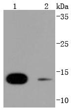
Fig1: Western blot analysis of SUMO4 on different lysates using anti-SUMO4 antibody at 1/1,000 dilution. Positive control: Lane 1: 293T Lane 2: JurkatApplication
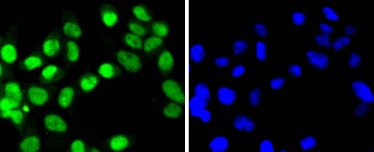
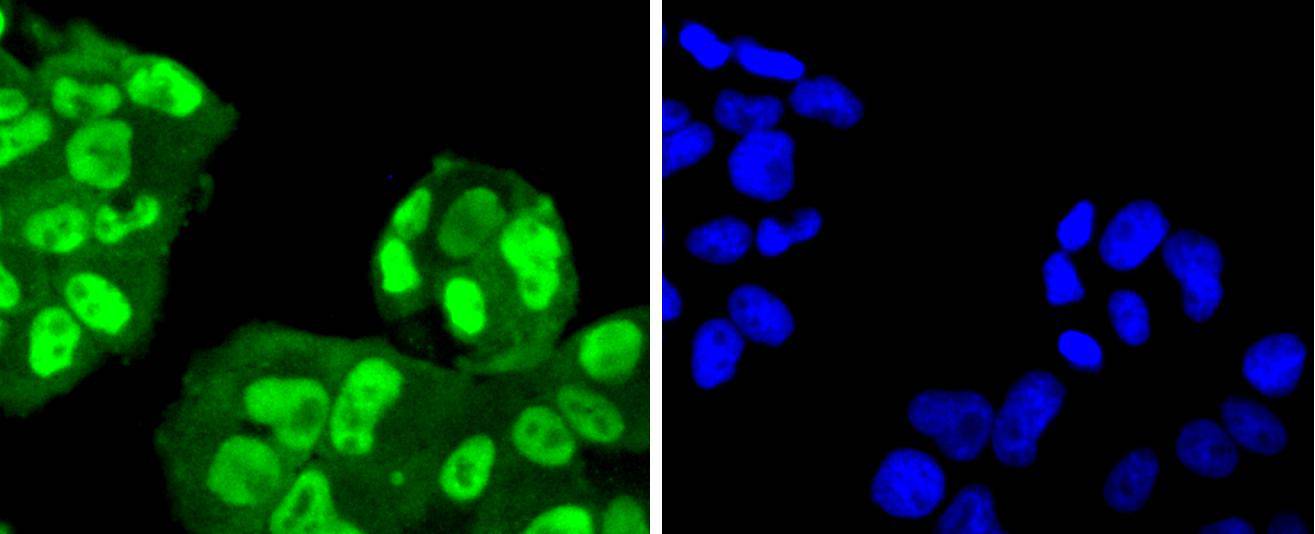
Fig2: ICC staining SUMO4 in 293 cells (green). The nuclear counter stain is DAPI (blue). Cells were fixed in paraformaldehyde, permeabilised with 0.25% Triton X100/PBS.Application
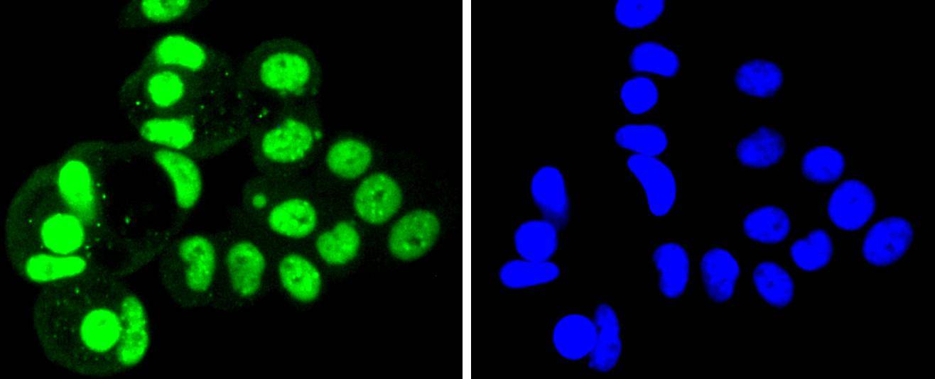
Fig3: ICC staining SUMO4 in MCF-7 cells (green). The nuclear counter stain is DAPI (blue). Cells were fixed in paraformaldehyde, permeabilised with 0.25% Triton X100/PBS.Application
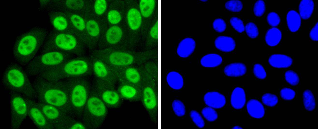
Fig4: ICC staining SUMO4 in HepG2 cells (green). The nuclear counter stain is DAPI (blue). Cells were fixed in paraformaldehyde, permeabilised with 0.25% Triton X100/PBS.Application
Fig5: ICC staining SUMO4 in RH-35 cells (green). The nuclear counter stain is DAPI (blue). Cells were fixed in paraformaldehyde, permeabilised with 0.25% Triton X100/PBS.Application
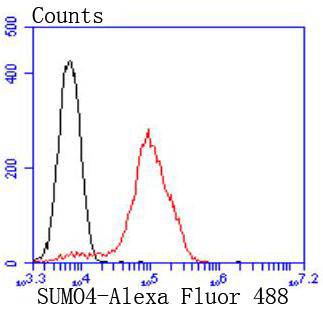
Fig6: Flow cytometric analysis of 293 cells with SUMO4 antibody at 1/50 dilution (red) compared with an unlabelled control (cells without incubation with primary antibody; black). Alexa Fluor 488-conjugated goat anti rabbit IgG was used as the secondary antibody| Product Name | SUMO4 [JJ085-01] |
|---|---|
| Antibody Type | Primary Antibodies |
| Antigen Alias | dJ281H8.4 antibody IDDM5 antibody Small ubiquitin like modifier 4 protein antibody Small ubiquitin-like protein 4 antibody Small ubiquitin-related modifier 4 antibody SMT3 suppressor of mif two 3 homolog 2 antibody SMT3 suppressor of mif two 3 homolog 4 (S. cerevisiae) antibody SMT3H4 antibody SUMO 4 antibody SUMO-4 antibody SUMO4 antibody SUMO4_HUMAN antibody |
| Product description | The small ubiquitin-related modifier (SUMO) proteins, which include SUMO-1, SUMO-2, SUMO-3 and SUMO-4, belong to the ubiquitin-like protein family. Like ubiquitin, the SUMO proteins are synthesized as precursor proteins that undergo processing before conjugation to target proteins. Ubiquitin and SUMO proteins utilize the E1, E2 and E3 cascade enzymes for conjugation. However, SUMO and ubiquitin differ with respect to targeting. Ubiquitination predominantly targets proteins for degradation, whereas sumoylation targets proteins for a variety of cellular processing, including nuclear transport, transcriptional regulation, apoptosis and protein stability. The unconjugated SUMO-1, SUMO-2, SUMO-3 and SUMO-4 proteins localize to the nucleus. In contrast to the other SUMO proteins, SUMO-4 seems to be insensitive to sentrin-specific proteases due to the presence of Pro-90, which may impair processing to mature form and conjugation to substrates. It is suggested that defects in the gene that encodes for the SUMO-4 protein may be involved in the pathogenesis of type I diabetes. |
| Immunogen | recombinant protein |
| Clonality | Monoclonal |
|---|---|
| Isotype | IgG |
| Host Species | Recombinant rabbit |
| Tested Applications | WBICC/IFIPFC |
| WB:1:1,000-1:2,000 FC:1:50-1:100 ICC:1:100-1:500 |
|
| Species Reactivity | HumanMouseRat |
| Concentration | 1mg/ml |
| Alternative Names | dJ281H8.4 antibody IDDM5 antibody Small ubiquitin like modifier 4 protein antibody Small ubiquitin-like protein 4 antibody Small ubiquitin-related modifier 4 antibody SMT3 suppressor of mif two 3 homolog 2 antibody SMT3 suppressor of mif two 3 homolog 4 (S. cerevisiae) antibody SMT3H4 antibody SUMO 4 antibody SUMO-4 antibody SUMO4 antibody SUMO4_HUMAN antibody |
|---|---|
| Molecular Weight(MW) | 11 kDa |
Application
Fig1: Western blot analysis of SUMO4 on different lysates using anti-SUMO4 antibody at 1/1,000 dilution. Positive control: Lane 1: 293T Lane 2: Jurkat
Application
Fig2: ICC staining SUMO4 in 293 cells (green). The nuclear counter stain is DAPI (blue). Cells were fixed in paraformaldehyde, permeabilised with 0.25% Triton X100/PBS.
Application
Fig3: ICC staining SUMO4 in MCF-7 cells (green). The nuclear counter stain is DAPI (blue). Cells were fixed in paraformaldehyde, permeabilised with 0.25% Triton X100/PBS.
Application
Fig4: ICC staining SUMO4 in HepG2 cells (green). The nuclear counter stain is DAPI (blue). Cells were fixed in paraformaldehyde, permeabilised with 0.25% Triton X100/PBS.
Application
Fig5: ICC staining SUMO4 in RH-35 cells (green). The nuclear counter stain is DAPI (blue). Cells were fixed in paraformaldehyde, permeabilised with 0.25% Triton X100/PBS.
Application
Fig6: Flow cytometric analysis of 293 cells with SUMO4 antibody at 1/50 dilution (red) compared with an unlabelled control (cells without incubation with primary antibody; black). Alexa Fluor 488-conjugated goat anti rabbit IgG was used as the secondary antibody| Positive Control | 293T, MCF-7, 293, HepG2, Jurkat, RH-35. |
|---|---|
| Application Notes | WB:1:1,000-1:2,000 FC:1:50-1:100 ICC:1:100-1:500 |
| Form | Liquid |
|---|---|
| Storage Instructions | Store at +4℃ after thawing. Aliquot store at -20℃ or -80℃. Avoid repeated freeze / thaw cycles. |
| Storage Buffer | 1*TBS (pH7.4), 1%BSA, 40%Glycerol. Preservative: 0.05% Sodium Azide. |


