Application
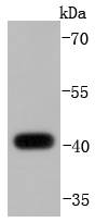
Fig1: Western blot analysis of DUSP1 on mouse spleen lysates using anti-DUSP1 antibody at 1/1,000 dilution.Application
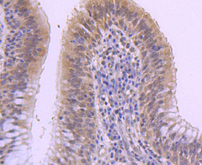
Fig2: Immunohistochemical analysis of paraffin-embedded human lung cancer tissue using anti-DUSP1 antibody. Counter stained with hematoxylin.Application
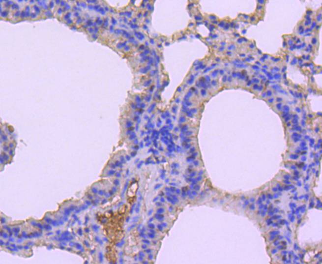
Fig3: Immunohistochemical analysis of paraffin-embedded mouse lung tissue using anti-DUSP1 antibody. Counter stained with hematoxylin.Application
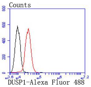
Fig4: Flow cytometric analysis of HepG2 cells with DUSP1 antibody at 1/50 dilution (red) compared with an unlabelled control (cells without incubation with primary antibody; black). Alexa Fluor 488-conjugated goat anti rabbit IgG was used as the secondary antibody| Product Name | DUSP1 [JJ0930] |
|---|---|
| Antibody Type | Primary Antibodies |
| Antigen Alias | CL 100 antibody CL100 antibody Dual Specificity Phosphatase 1 antibody Dual specificity protein phosphatase 1 antibody Dual specificity protein phosphatase hVH1 antibody DUS1_HUMAN antibody DUSP 1 antibody Dusp1 antibody HVH1 antibody MAP kinase phosphatase 1 antibody Mitogen-activated protein kinase phosphatase 1 antibody MKP-1 antibody MKP1 antibody Protein tyrosine phosphatase CL100 antibody Protein-tyrosine phosphatase CL100 antibody PTPN10 antibody Serine/threonine specific protein phosphatase antibody VH1 antibody |
| Product description | A key element in the pathway involved in the transduction of signals from activated protein-tyrosine kinase transmembrane receptors has been identified as the family of mitogen-activated protein kinases (MAP kinases). The most well known of these Ser/Thr kinases are ERK 1 and ERK 2. Mitogenic stimulation of cells triggers the activation of MAP kinases through phosphorylation of both tyrosyl (Y185) and threonyl (T183) residues. Phosphorylation of the T183 and Y185 ERK regulatory site is mediated by MAP kinase (MEK), which in turn is regulated by the proto-oncogene product Raf. Two highly related phosphatases, designated MKP-1 and MKP-2, exhibit 59% sequence identity at the amino acid level and oppose the action of MEK by downregulating the kinase activity of ERK 1 and ERK 2. MAP kinase phosphatase-1 and -2 proteins function by dephosphorylating ERK 1 and ERK 2 at their T-E-Y regulatory motif. An additional phosphatase encoded by the DUSP2 gene, designated PAC-1, also functions to downregulate ERK 1 and ERK 2 kinase activity. PAC-1 is a nuclear protein whose expression is strongly induced in response to mitogen. |
| Immunogen | recombinant protein |
| Clonality | Monoclonal |
|---|---|
| Isotype | IgG |
| Host Species | Recombinant rabbit |
| Tested Applications | WBIHCIPFC |
| WB:1:1,000 FC:1:50-1:100 IHC:1:50-1:200 |
|
| Species Reactivity | HumanMouseRat |
| Concentration | 1mg/ml |
| Alternative Names | CL 100 antibody CL100 antibody Dual Specificity Phosphatase 1 antibody Dual specificity protein phosphatase 1 antibody Dual specificity protein phosphatase hVH1 antibody DUS1_HUMAN antibody DUSP 1 antibody Dusp1 antibody HVH1 antibody MAP kinase phosphatase 1 antibody Mitogen-activated protein kinase phosphatase 1 antibody MKP-1 antibody MKP1 antibody Protein tyrosine phosphatase CL100 antibody Protein-tyrosine phosphatase CL100 antibody PTPN10 antibody Serine/threonine specific protein phosphatase antibody VH1 antibody |
|---|---|
| Molecular Weight(MW) | 40 kDa |
| Cellular Localization | Nucleus. |
Application
Fig1: Western blot analysis of DUSP1 on mouse spleen lysates using anti-DUSP1 antibody at 1/1,000 dilution.
Application
Fig2: Immunohistochemical analysis of paraffin-embedded human lung cancer tissue using anti-DUSP1 antibody. Counter stained with hematoxylin.
Application
Fig3: Immunohistochemical analysis of paraffin-embedded mouse lung tissue using anti-DUSP1 antibody. Counter stained with hematoxylin.
Application
Fig4: Flow cytometric analysis of HepG2 cells with DUSP1 antibody at 1/50 dilution (red) compared with an unlabelled control (cells without incubation with primary antibody; black). Alexa Fluor 488-conjugated goat anti rabbit IgG was used as the secondary antibody| Positive Control | Human lung cancer tissue, mouse spleen tissue, mouse lung tissue. |
|---|---|
| Application Notes | WB:1:1,000 FC:1:50-1:100 IHC:1:50-1:200 |
| Form | Liquid |
|---|---|
| Storage Instructions | Store at +4℃ after thawing. Aliquot store at -20℃ or -80℃. Avoid repeated freeze / thaw cycles. |
| Storage Buffer | 1*TBS (pH7.4), 1%BSA, 40%Glycerol. Preservative: 0.05% Sodium Azide. |


