WB
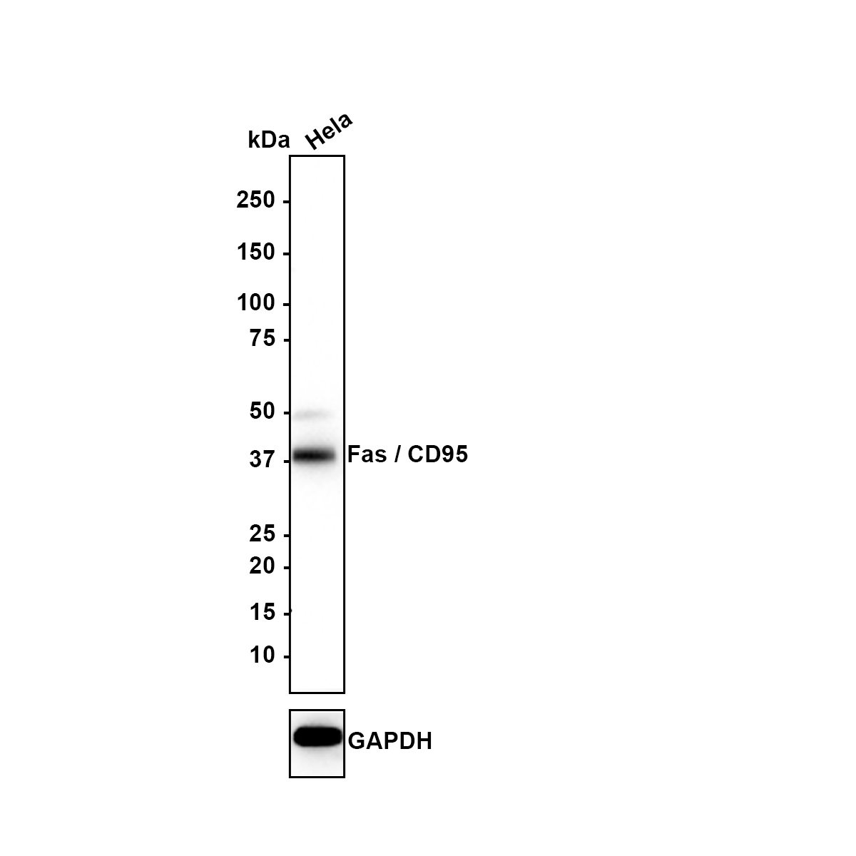
Western blot analysis of Fas / CD95 on HeLa (Human cervical adenocarcinoma cell) lysates with Rabbit anti-Fas / CD95 antibody at 1/1,000 dilution. Lysates/proteins at 15 µg/Lane. Exposure time: 3 minutes; Blocking: 5% NFDM/TBST, 1 hour at room temperature, Primary antibody: 1/1,000 in primary antibody dilution buffer, overnight at 4 ℃, Secondary antibody: Goat anti-Rabbit IgG-HRP, 1/50,000 in 5% NFDM/TBST, 1 hour at room temperature.IHC
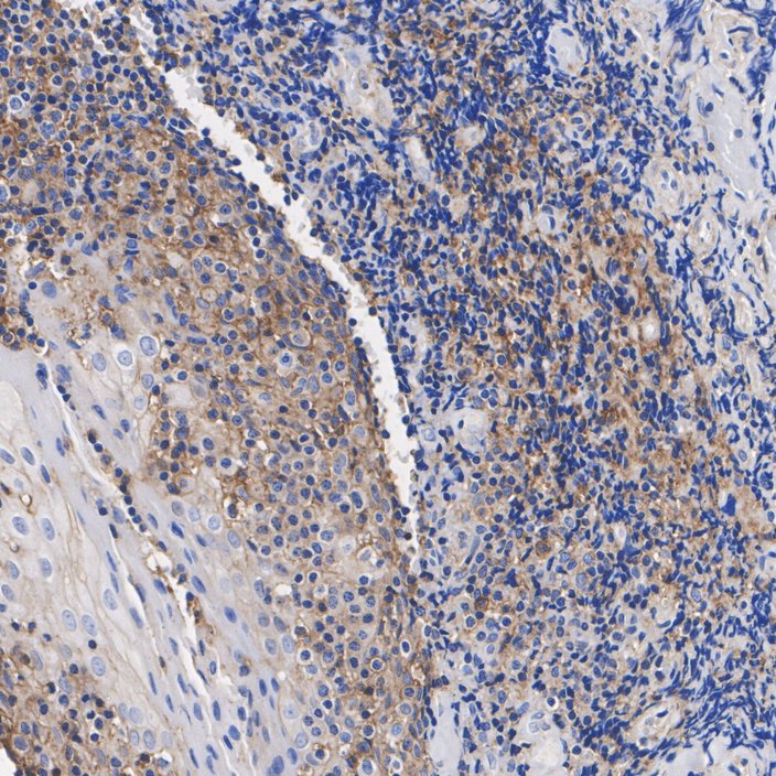
Immunohistochemical analysis of paraffin-embedded human tonsil tissue with Rabbit anti-Fas / CD95 antibody at 1/200 dilution. The section was pre-treated using heat mediated antigen retrieval with Tris-EDTA buffer (pH 9.0) for 20 minutes. The tissues were blocked in 1% BSA for 20 minutes at room temperature, washed with ddH2O and PBS, and then probed with the primary antibody at 1/200 dilution for 1 hour at room temperature. The detection was performed using an HRP conjugated compact polymer system. DAB was used as the chromogen. Tissues were counterstained with hematoxylin and mounted with DPX.IHC
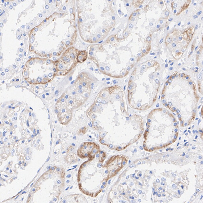
Immunohistochemical analysis of paraffin-embedded human kidney tissue with Rabbit anti-Fas / CD95 antibody at 1/1,000 dilution. The section was pre-treated using heat mediated antigen retrieval with Tris-EDTA buffer (pH 9.0) for 20 minutes. The tissues were blocked in 1% BSA for 20 minutes at room temperature, washed with ddH2O and PBS, and then probed with the primary antibody at 1/1,000 dilution for 1 hour at room temperature. The detection was performed using an HRP conjugated compact polymer system. DAB was used as the chromogen. Tissues were counterstained with hematoxylin and mounted with DPX.| Product Name | Fas / CD95 Recombinant Rabbit Monoclonal Antibody |
|---|---|
| Antibody Type | Primary Antibodies |
| Product description | Cytotoxic T lymphocyte (CTL)-mediated cytotoxicity constitutes an important component of specific effector mechanisms in immuno-surveillance against virus-infected or transformed cells. Two mechanisms appear to account for this activity, one of which is the perforin-based process. Independently, a FAS-based mechanism involves the transducing molecule FAS (also designated APO-1) and its ligand (FAS-L). The human FAS protein is a cell surface glycoprotein that belongs to a family of receptors that includes CD40, nerve growth factor receptors and tumor necrosis factor receptors. The FAS antigen is expressed on a broad range of lymphoid cell lines, certain of which undergo apoptosis in response to treatment with antibody to FAS. These findings strongly imply that targeted cell death is potentially mediated by the intercellular interactions of FAS with its ligand or effectors, and that FAS may be critically involved in CTL-mediated cytotoxicity. |
| Immunogen | Recombinant protein within Human Fas aa 130-335 / 335. |
| Clonality | Monoclonal |
|---|---|
| Isotype | IgG |
| Host Species | Recombinant rabbit |
| Tested Applications | IHCWB |
| WB:1:500-1:2000 IHC:1:50-1:200 |
|
| Species Reactivity | Human |
| Concentration | 1mg/ml |
| Purification | Protein A |
| Alternative Names | ALPS 1A antibody
ALPS1A antibody
APO 1 antibody
Apo 1 antigen antibody
APO 1 cell surface antigen antibody
Apo-1 antigen antibody
APO1 antibody
Apo1 antigen antibody
APO1 cell surface antigen antibody
Apoptosis antigen 1 antibody
Apoptosis mediating surface antigen FAS antibody
Apoptosis-mediating surface antigen FAS antibody
APT 1 antibody
APT1 antibody
CD 95 antibody
CD 95 antigen antibody
CD95 antibody
CD95 antigen antibody
Delta Fas antibody
Delta Fas/APO 1/CD95 antibody
Delta Fas/APO1/CD95 antibody
Fas (TNF receptor superfamily member 6) antibody FAS 1 antibody FAS 827dupA antibody Fas AMA antibody Fas antibody FAS Antigen antibody Fas cell surface death receptor antibody FAS1 antibody FASLG receptor antibody FASTM antibody sFAS antibody Surface antigen APO1 antibody TNF receptor superfamily member 6 antibody TNFRSF 6 antibody TNFRSF6 antibody TNR6_HUMAN antibody Tumor necrosis factor receptor superfamily member 6 antibody |
|---|---|
| Molecular Weight(MW) | 37 kDa |
| Cellular Localization | Cell membrane, Secreted. |
| SwissProt ID | P25445 |
|---|
WB
Western blot analysis of Fas / CD95 on HeLa (Human cervical adenocarcinoma cell) lysates with Rabbit anti-Fas / CD95 antibody at 1/1,000 dilution. Lysates/proteins at 15 µg/Lane. Exposure time: 3 minutes; Blocking: 5% NFDM/TBST, 1 hour at room temperature, Primary antibody: 1/1,000 in primary antibody dilution buffer, overnight at 4 ℃, Secondary antibody: Goat anti-Rabbit IgG-HRP, 1/50,000 in 5% NFDM/TBST, 1 hour at room temperature.
IHC
Immunohistochemical analysis of paraffin-embedded human tonsil tissue with Rabbit anti-Fas / CD95 antibody at 1/200 dilution. The section was pre-treated using heat mediated antigen retrieval with Tris-EDTA buffer (pH 9.0) for 20 minutes. The tissues were blocked in 1% BSA for 20 minutes at room temperature, washed with ddH2O and PBS, and then probed with the primary antibody at 1/200 dilution for 1 hour at room temperature. The detection was performed using an HRP conjugated compact polymer system. DAB was used as the chromogen. Tissues were counterstained with hematoxylin and mounted with DPX.
IHC
Immunohistochemical analysis of paraffin-embedded human kidney tissue with Rabbit anti-Fas / CD95 antibody at 1/1,000 dilution. The section was pre-treated using heat mediated antigen retrieval with Tris-EDTA buffer (pH 9.0) for 20 minutes. The tissues were blocked in 1% BSA for 20 minutes at room temperature, washed with ddH2O and PBS, and then probed with the primary antibody at 1/1,000 dilution for 1 hour at room temperature. The detection was performed using an HRP conjugated compact polymer system. DAB was used as the chromogen. Tissues were counterstained with hematoxylin and mounted with DPX.| Application Notes | WB:1:500-1:2000 IHC:1:50-1:200 |
|---|
| Form | Liquid |
|---|---|
| Storage Instructions | Store at +4℃ after thawing. Aliquot store at -20℃ or -80℃. Avoid repeated freeze / thaw cycles. |
| Storage Buffer | 1*TBS (pH7.4), 1%BSA, 40%Glycerol. Preservative: 0.05% Sodium Azide. |


