Application
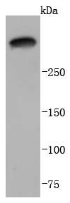
Fig1: Western blot analysis of Laminin 5 alpha 3 on A431 cells lysates using anti-Laminnin 5 alpha 3 antibody at 1/1,000 dilution.Application
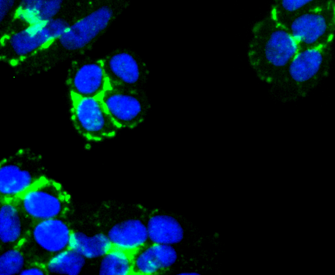
Fig2: ICC staining Laminin 5 alpha 3 in Hela cells (green). The nuclear counter stain is DAPI (blue). Cells were fixed in paraformaldehyde, permeabilised with 0.25% Triton X100/PBS.Application
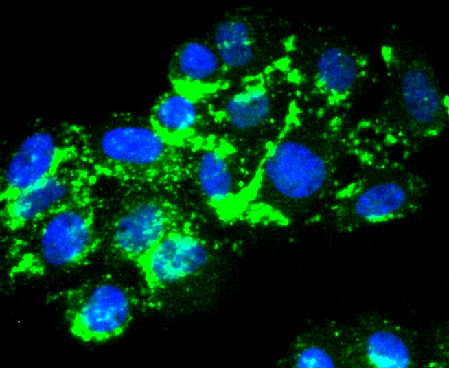
Fig3: ICC staining Laminin 5 alpha 3 in HepG2 cells (green). The nuclear counter stain is DAPI (blue). Cells were fixed in paraformaldehyde, permeabilised with 0.25% Triton X100/PBS.| Product Name | Laminin 5 alpha 3 [JJ0957] |
|---|---|
| Antibody Type | Primary Antibodies |
| Antigen Alias | E170 antibody Epiligrin 170 kDa subunit antibody Epiligrin subunit alpha antibody Kalinin subunit alpha antibody LAMA3 antibody LAMA3A antibody Laminin 5 alpha3 antibody Laminin A3 antibody laminin alpha 3 antibody laminin alpha3 antibody Laminin subunit alpha-3 antibody Laminin-5 subunit alpha antibody Laminin-6 subunit alpha antibody Laminin-7 subunit alpha antibody laminin5 alpha 3 antibody Laminin5 alpha3 antibody LAMNA antibody LOCS antibody Nicein subunit alpha antibody |
| Product description | Laminins are heterotrimeric, noncollagenous glycoproteins composed of alpha, beta, and gamma chains. Through interactions with integrins, dystroglycan and other receptors, laminins contribute to cell differentiation, cell shape and migration, and maintenance of tissue phenotypes and survival. Laminin alpha 3/Laminin-5, also known as epiligrin, includes alpha 3, beta 3, and gamma 2 subunits. It is abundant in transitional epithelium, stratified squamous epithelia, lung mucosa and other epithelial glands and contributes to initiation and maintenance of epithelial cell anchorage to the underlying connective tissue. Within aa 21?1713 of the alpha 3 subunit, human and mouse share 77% amino acid sequence identity. |
| Immunogen | recombinant protein |
| Clonality | Monoclonal |
|---|---|
| Isotype | IgG |
| Host Species | Recombinant rabbit |
| Tested Applications | WBICC/IF |
| WB:1:1,000-1:2,000 ICC:1:100-1:500 |
|
| Species Reactivity | Human |
| Concentration | 1mg/ml |
| Alternative Names | E170 antibody Epiligrin 170 kDa subunit antibody Epiligrin subunit alpha antibody Kalinin subunit alpha antibody LAMA3 antibody LAMA3A antibody Laminin 5 alpha3 antibody Laminin A3 antibody laminin alpha 3 antibody laminin alpha3 antibody Laminin subunit alpha-3 antibody Laminin-5 subunit alpha antibody Laminin-6 subunit alpha antibody Laminin-7 subunit alpha antibody laminin5 alpha 3 antibody Laminin5 alpha3 antibody LAMNA antibody LOCS antibody Nicein subunit alpha antibody |
|---|---|
| Molecular Weight(MW) | 367 kDa |
| Cellular Localization | Secreted. |
| SwissProt ID | Q16787 |
|---|
Application
Fig1: Western blot analysis of Laminin 5 alpha 3 on A431 cells lysates using anti-Laminnin 5 alpha 3 antibody at 1/1,000 dilution.
Application
Fig2: ICC staining Laminin 5 alpha 3 in Hela cells (green). The nuclear counter stain is DAPI (blue). Cells were fixed in paraformaldehyde, permeabilised with 0.25% Triton X100/PBS.
Application
Fig3: ICC staining Laminin 5 alpha 3 in HepG2 cells (green). The nuclear counter stain is DAPI (blue). Cells were fixed in paraformaldehyde, permeabilised with 0.25% Triton X100/PBS.| Positive Control | A431, Hela, HepG2. |
|---|---|
| Application Notes | WB:1:1,000-1:2,000 ICC:1:100-1:500 |
| Form | Liquid |
|---|---|
| Storage Instructions | Store at +4℃ after thawing. Aliquot store at -20℃ or -80℃. Avoid repeated freeze / thaw cycles. |
| Storage Buffer | 1*TBS (pH7.4), 1%BSA, 40%Glycerol. Preservative: 0.05% Sodium Azide. |


