Application
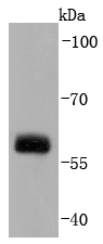
Fig1: Western blot analysis of Src on HUVEC cells lysates using anti-Src antibody at 1/1,000 dilution.Application
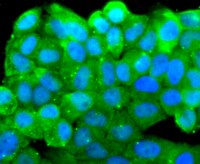
Fig2: ICC staining Src in Hela cells (green). The nuclear counter stain is DAPI (blue). Cells were fixed in paraformaldehyde, permeabilised with 0.25% Triton X100/PBS.Application
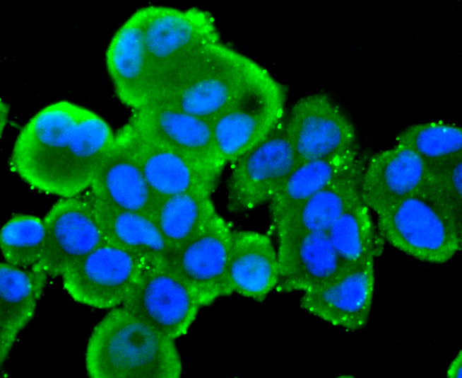
Fig3: ICC staining Src in MCF-7 cells (green). The nuclear counter stain is DAPI (blue). Cells were fixed in paraformaldehyde, permeabilised with 0.25% Triton X100/PBS.Application
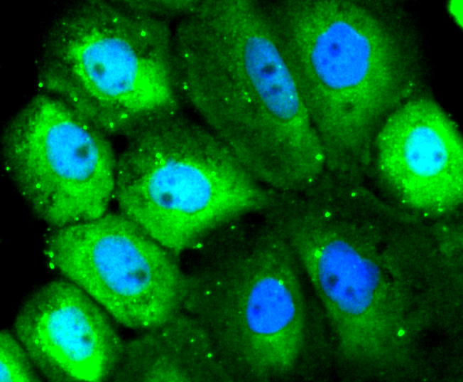
Fig4: ICC staining Src in A431 cells (green). The nuclear counter stain is DAPI (blue). Cells were fixed in paraformaldehyde, permeabilised with 0.25% Triton X100/PBS.Application
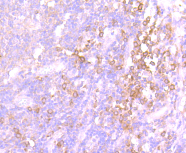
Fig5: Immunohistochemical analysis of paraffin-embedded human tonsil tissue using anti-Src antibody. Counter stained with hematoxylin.Application
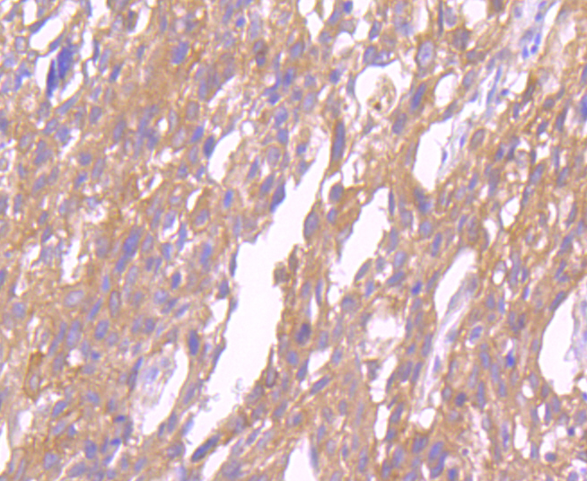
Fig6: Immunohistochemical analysis of paraffin-embedded human breast carcinoma tissue using anti-Src antibody. Counter stained with hematoxylin.Application
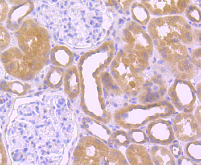
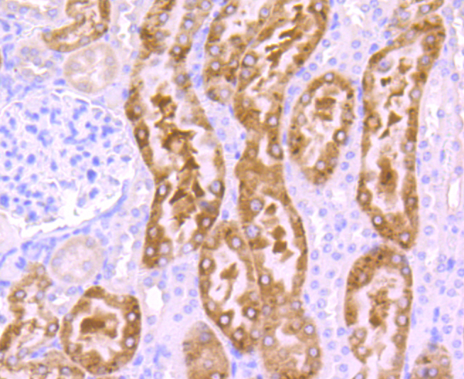
Fig7: Immunohistochemical analysis of paraffin-embedded human kidney tissue using anti-Src antibody. Counter stained with hematoxylin.Application
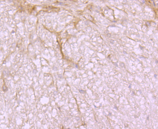
Fig8: Immunohistochemical analysis of paraffin-embedded rat brain tissue using anti-Src antibody. Counter stained with hematoxylin.Application
Fig9: Immunohistochemical analysis of paraffin-embedded mouse kidney tissue using anti-Src antibody. Counter stained with hematoxylin.Application
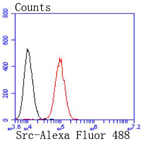
Fig10: Flow cytometric analysis of NIH/3T3 cells with Src antibody at 1/50 dilution (red) compared with an unlabelled control (cells without incubation with primary antibody; black). Alexa Fluor 488-conjugated goat anti rabbit IgG was used as the secondary antibody| Product Name | Src [JF0947] |
|---|---|
| Antibody Type | Primary Antibodies |
| Antigen Alias | ASV antibody Avian sarcoma virus antibody c SRC antibody CDNA FLJ14219 fis clone NT2RP3003800 highly similar to Rattus norvegicus tyrosine protein kinase pp60 c src mRNA antibody cSrc antibody EC 2.7.10.2 antibody Neuronal CSRC tyrosine specific protein kinase antibody Neuronal SRC antibody Oncogene SRC antibody OTTHUMP00000174476 antibody OTTHUMP00000174477 antibody p60 Src antibody p60-Src antibody p60Src antibody pp60c src antibody pp60c-src antibody pp60csrc antibody Proto oncogene tyrosine protein kinase Src antibody Proto-oncogene c-Src antibody Proto-oncogene tyrosine-protein kinase Src antibody Protooncogene SRC antibody Protooncogene SRC Rous sarcoma antibody Src antibody SRC Oncogene antibody SRC proto oncogene non receptor tyrosine kinase antibody SRC_HUMAN antibody SRC1 antibody Tyrosine kinase pp60c src antibody Tyrosine protein kinase SRC 1 antibody Tyrosine protein kinase SRC1 antibody v src avian sarcoma (Schmidt Ruppin A2) viral oncogene homolog antibody V src sarcoma (Schmidt Ruppin A 2) viral oncogene homolog (avian) antibody v src sarcoma (Schmidt Ruppin A 2) viral oncogene homolog avian antibody |
| Product description | The major translational products of the Src gene family are membrane-associated tyrosine protein kinases that lack transmembrane and external amino acid sequences. By virtue of their common structural motifs, the Src family is composed of nine members in vertebrates, including c-Src, c-Yes, Fgr, Yrk, Fyn, Lyn, Hck, Lck and Blk. Src family kinases, which contain an amino-terminal cell membrane anchor followed by SH3 and SH2 domains, transduce signals that are involved in the control of a variety of cellular processes, including proliferation, differentiation, motility and adhesion. Src family members are normally maintained in an inactive state and can be activated transiently during cellular events such as mitosis. Different subcellular locations of Src family kinases may be important for the regulation of specific cellular processes, such as mitogenesis, cytoskeletal organization and membrane trafficking. c-Src (also designated pp60Src, Src p60 and proto-oncogene tyrosine protein kinase Src) is expressed in a broad range of tissue and cell types, although the highest levels of c-Src are detected in neuronal tissues and platelets. c-Src may play a role in events associated with both neuronal differentiation and maintenance of mature neuronal cell functions. |
| Immunogen | recombinant protein |
| Clonality | Monoclonal |
|---|---|
| Isotype | IgG |
| Host Species | Recombinant rabbit |
| Tested Applications | WBICC/IFIHCFC |
| WB:1:1,000-1:5,000 ICC:1:100-1:500 IHC:1:50-1:200 FC:1:50-1:100 |
|
| Species Reactivity | HumanMouseRat |
| Concentration | 1mg/ml |
| Alternative Names | ASV antibody Avian sarcoma virus antibody c SRC antibody CDNA FLJ14219 fis clone NT2RP3003800 highly similar to Rattus norvegicus tyrosine protein kinase pp60 c src mRNA antibody cSrc antibody EC 2.7.10.2 antibody Neuronal CSRC tyrosine specific protein kinase antibody Neuronal SRC antibody Oncogene SRC antibody OTTHUMP00000174476 antibody OTTHUMP00000174477 antibody p60 Src antibody p60-Src antibody p60Src antibody pp60c src antibody pp60c-src antibody pp60csrc antibody Proto oncogene tyrosine protein kinase Src antibody Proto-oncogene c-Src antibody Proto-oncogene tyrosine-protein kinase Src antibody Protooncogene SRC antibody Protooncogene SRC Rous sarcoma antibody Src antibody SRC Oncogene antibody SRC proto oncogene non receptor tyrosine kinase antibody SRC_HUMAN antibody SRC1 antibody Tyrosine kinase pp60c src antibody Tyrosine protein kinase SRC 1 antibody Tyrosine protein kinase SRC1 antibody v src avian sarcoma (Schmidt Ruppin A2) viral oncogene homolog antibody V src sarcoma (Schmidt Ruppin A 2) viral oncogene homolog (avian) antibody v src sarcoma (Schmidt Ruppin A 2) viral oncogene homolog avian antibody |
|---|---|
| Molecular Weight(MW) | 60 kDa |
| Cellular Localization | Cell membrane, Mitochondrion inner membrane, Nucleus, Cytoplasm. |
Application
Fig1: Western blot analysis of Src on HUVEC cells lysates using anti-Src antibody at 1/1,000 dilution.
Application
Fig2: ICC staining Src in Hela cells (green). The nuclear counter stain is DAPI (blue). Cells were fixed in paraformaldehyde, permeabilised with 0.25% Triton X100/PBS.
Application
Fig3: ICC staining Src in MCF-7 cells (green). The nuclear counter stain is DAPI (blue). Cells were fixed in paraformaldehyde, permeabilised with 0.25% Triton X100/PBS.
Application
Fig4: ICC staining Src in A431 cells (green). The nuclear counter stain is DAPI (blue). Cells were fixed in paraformaldehyde, permeabilised with 0.25% Triton X100/PBS.
Application
Fig5: Immunohistochemical analysis of paraffin-embedded human tonsil tissue using anti-Src antibody. Counter stained with hematoxylin.
Application
Fig6: Immunohistochemical analysis of paraffin-embedded human breast carcinoma tissue using anti-Src antibody. Counter stained with hematoxylin.
Application
Fig7: Immunohistochemical analysis of paraffin-embedded human kidney tissue using anti-Src antibody. Counter stained with hematoxylin.
Application
Fig8: Immunohistochemical analysis of paraffin-embedded rat brain tissue using anti-Src antibody. Counter stained with hematoxylin.
Application
Fig9: Immunohistochemical analysis of paraffin-embedded mouse kidney tissue using anti-Src antibody. Counter stained with hematoxylin.
Application
Fig10: Flow cytometric analysis of NIH/3T3 cells with Src antibody at 1/50 dilution (red) compared with an unlabelled control (cells without incubation with primary antibody; black). Alexa Fluor 488-conjugated goat anti rabbit IgG was used as the secondary antibody| Positive Control | HUVEC, MCF-7, Hela, A431, rat brain tissue, mouse kidney tissue, human tonsil tissue, human breast carcinoma tissue, human kidney tissue. |
|---|---|
| Application Notes | WB:1:1,000-1:5,000 ICC:1:100-1:500 IHC:1:50-1:200 FC:1:50-1:100 |
| Form | Liquid |
|---|---|
| Storage Instructions | Store at +4℃ after thawing. Aliquot store at -20℃ or -80℃. Avoid repeated freeze / thaw cycles. |
| Storage Buffer | 1*TBS (pH7.4), 1%BSA, 40%Glycerol. Preservative: 0.05% Sodium Azide. |


