WB
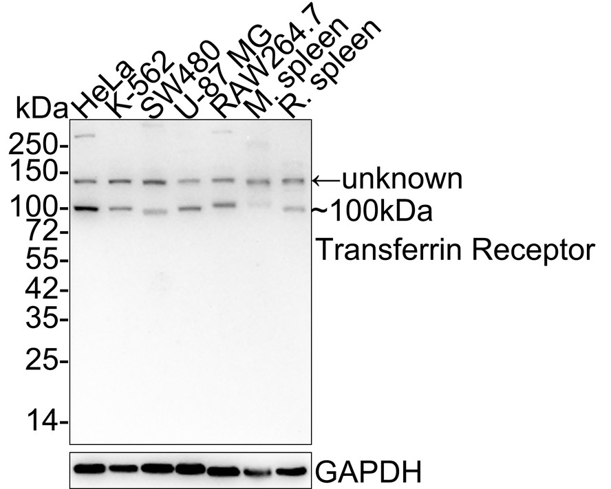
Western blot analysis of Transferrin Receptor (CD71) on different lysates with Rabbit anti-Transferrin Receptor (CD71) antibody at 1/2,000 dilution. Lane 1: HeLa cell lysate, Lane 2: K-562 cell lysate, Lane 3: SW480 cell lysate, Lane 4: U-87 MG cell lysate, Lane 5: RAW264.7 cell lysate, Lane 6: Mouse spleen tissue lysate, Lane 7: Rat spleen tissue lysate, Lysates/proteins at 20 µg/Lane. Exposure time: 5 minutes; 4-20% SDS-PAGE gel. Proteins were transferred to a PVDF membrane and blocked with 5% NFDM/TBST for 1 hour at room temperature. The primary antibody at 1/2,000 dilution was used in 5% NFDM/TBST at 4℃ overnight. Goat Anti-Rabbit IgG - HRP Secondary Antibody at 1:50,000 dilution was used for 1 hour at room temperature.FC
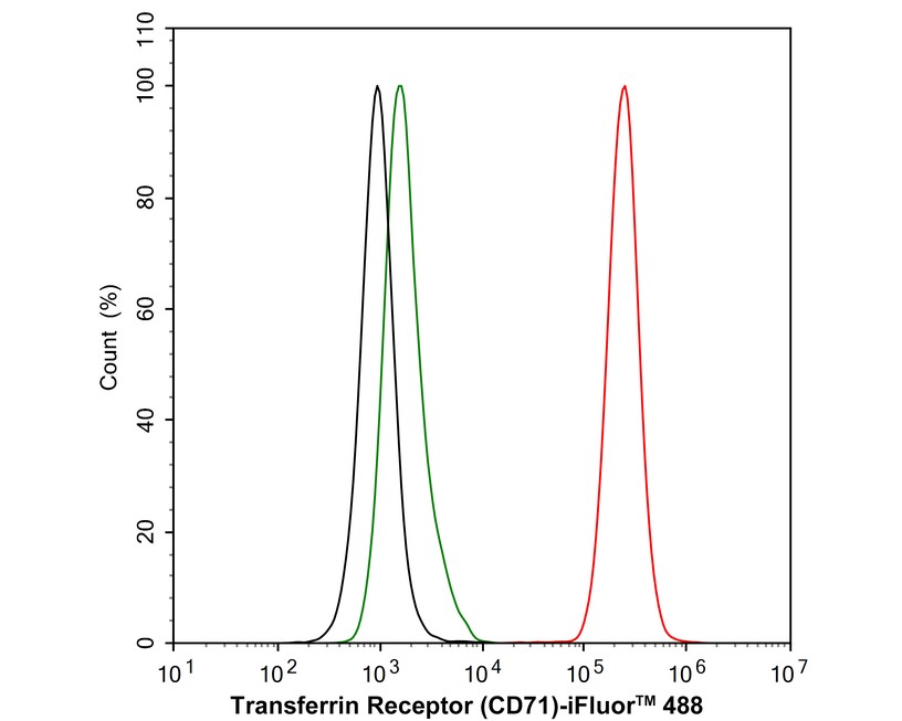
Flow cytometric analysis of HeLa cells labeling Transferrin Receptor (CD71). Cells were fixed and permeabilized. Then stained with the primary antibody (1μg/mL) (red) compared with Rabbit IgG Isotype Control (green). After incubation of the primary antibody at +4℃ for an hour, the cells were stained with a 488 conjugate-Goat anti-Rabbit IgG Secondary antibody at 1/1,000 dilution for 30 minutes at +4℃. Unlabelled sample was used as a control (cells without incubation with primary antibody; black).| Product Name | Transferrin Receptor (CD71) Recombinant Rabbit Monoclonal Antibody |
|---|---|
| Antibody Type | Primary Antibodies |
| Product description | CD71, also known as the transferrin receptor (TFR), is a type II membrane glycoprotein that exists as a disulfide-linked homodimer of two identical subunits. CD71 binds to two molecules of transferrin and a serum iron-transport protein, and directs the cellular uptake of iron via receptor-mediated endocytosis. CD71 is expressed, typically at high levels, on all proliferating cells, reticulocytes and erythroid precursors. It is not expressed on resting leukocytes, but is upregulated upon activation of lymphocytes, monocytes and macrophages. CD71 is also found on most dividing cells and on brain endothelium. A second transferrin receptor, TFR2, also mediates the uptake of transferrin-bound iron. TFR2 is a two-subunit homodimer and is highly expressed in liver as well as in hepatocytes and erythroid precursors. Mutations in the TFR2 gene result in hereditary hemochromatosis type III (HFE3), an iron overloading disorder predominant in Caucasians. |
| Immunogen | Synthetic peptide within Human CD71 aa 22-60 / 760. |
| Clonality | Monoclonal |
|---|---|
| Isotype | IgG |
| Host Species | Recombinant rabbit |
| Tested Applications | FCIPWB |
| WB:1:1000-1:10000 FC:1:1000 IP: Use at an assay dependent concentration. |
|
| Species Reactivity | HumanMouseRat |
| Concentration | 1mg/ml |
| Purification | Protein A |
| Alternative Names | CD 71 antibody
CD71 antibody
CD71 antigen antibody
IMD46 antibody
OTTHUMP00000208523 antibody
OTTHUMP00000208524 antibody
OTTHUMP00000208525 antibody
p90 antibody
sTfR antibody
T9 antibody
TFR 1 antibody
TfR antibody
TfR1 antibody
TFR1_HUMAN antibody
TFRC antibody
TR antibody
Transferrin receptor (p90 CD71) antibody
Transferrin receptor protein 1 serum form antibody Trfr antibody |
|---|---|
| Molecular Weight(MW) | 85 kDa(Observed band size: 100 kDa) |
| Cellular Localization | Cell membrane, Melanosome, Secreted. |
WB
Western blot analysis of Transferrin Receptor (CD71) on different lysates with Rabbit anti-Transferrin Receptor (CD71) antibody at 1/2,000 dilution. Lane 1: HeLa cell lysate, Lane 2: K-562 cell lysate, Lane 3: SW480 cell lysate, Lane 4: U-87 MG cell lysate, Lane 5: RAW264.7 cell lysate, Lane 6: Mouse spleen tissue lysate, Lane 7: Rat spleen tissue lysate, Lysates/proteins at 20 µg/Lane. Exposure time: 5 minutes; 4-20% SDS-PAGE gel. Proteins were transferred to a PVDF membrane and blocked with 5% NFDM/TBST for 1 hour at room temperature. The primary antibody at 1/2,000 dilution was used in 5% NFDM/TBST at 4℃ overnight. Goat Anti-Rabbit IgG - HRP Secondary Antibody at 1:50,000 dilution was used for 1 hour at room temperature.
FC
Flow cytometric analysis of HeLa cells labeling Transferrin Receptor (CD71). Cells were fixed and permeabilized. Then stained with the primary antibody (1μg/mL) (red) compared with Rabbit IgG Isotype Control (green). After incubation of the primary antibody at +4℃ for an hour, the cells were stained with a 488 conjugate-Goat anti-Rabbit IgG Secondary antibody at 1/1,000 dilution for 30 minutes at +4℃. Unlabelled sample was used as a control (cells without incubation with primary antibody; black).| Application Notes | WB:1:1000-1:10000 FC:1:1000 IP: Use at an assay dependent concentration. |
|---|
| Form | Liquid |
|---|---|
| Storage Instructions | Store at +4℃ after thawing. Aliquot store at -20℃ or -80℃. Avoid repeated freeze / thaw cycles. |
| Storage Buffer | 1*TBS (pH7.4), 1%BSA, 40%Glycerol. Preservative: 0.05% Sodium Azide. |


