WB
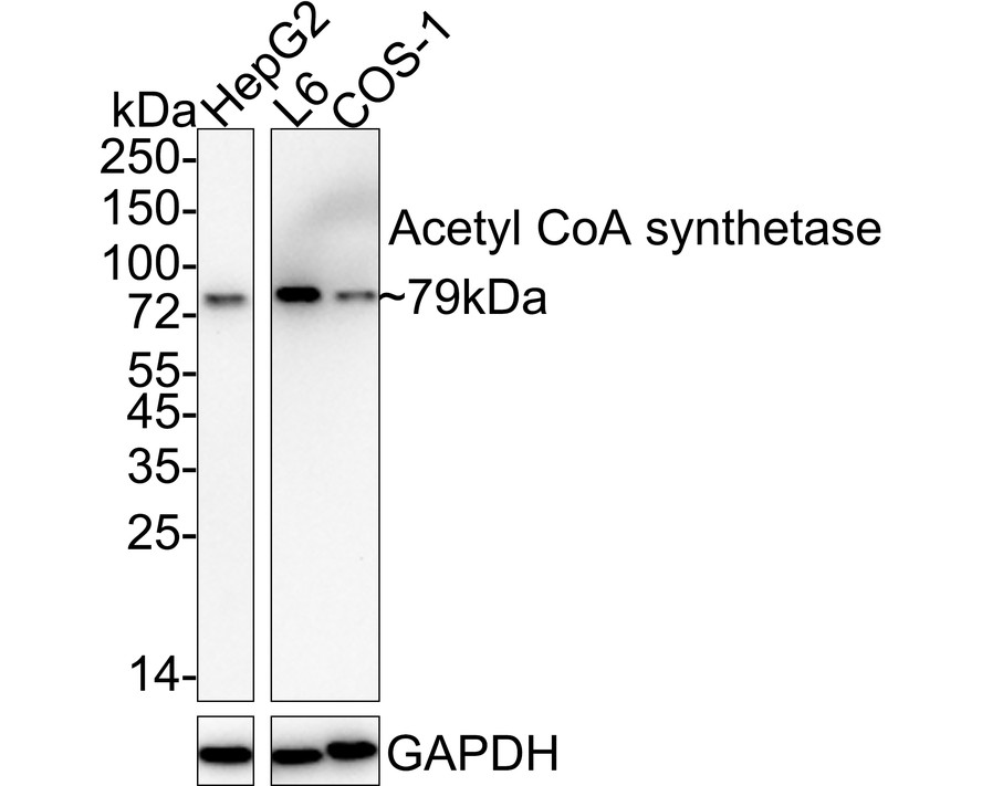
Western blot analysis of Acetyl CoA synthetase on different lysates with Rabbit anti-Acetyl CoA synthetase antibody at 1/1,000 dilution. Lane 1: HepG2 cell lysate, Lane 2: L6 cell lysate, Lane 3: COS-1 cell lysate, Lysates/proteins at 20 µg/Lane. Exposure time: 3 minutes; 4-20% SDS-PAGE gel. Proteins were transferred to a PVDF membrane and blocked with 5% NFDM/TBST for 1 hour at room temperature. The primary antibody at 1/1,000 dilution was used in 5% NFDM/TBST at 4℃ overnight. Goat Anti-Rabbit IgG - HRP Secondary Antibody at 1/50,000 dilution was used for 1 hour at room temperature.WB
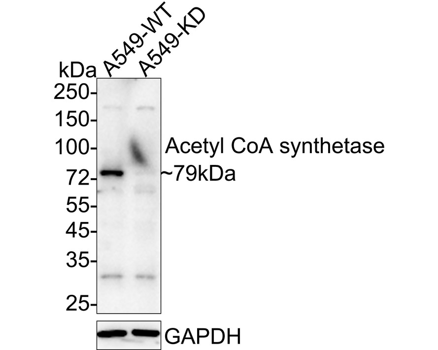
Western blot analysis of Acetyl CoA synthetase on different lysates with Rabbit anti-Acetyl CoA synthetase antibody at 1/2,000 dilution. Lane 1: A549-si NT cell lysate, Lane 2: A549-si Acetyl CoA synthetase cell lysate, Lysates/proteins at 10 µg/Lane. Exposure time: 3 minutes; 4-20% SDS-PAGE gel. Proteins were transferred to a PVDF membrane and blocked with 5% NFDM/TBST for 1 hour at room temperature. The primary antibody at 1/2,000 dilution was used in 5% NFDM/TBST at 4℃ overnight. Goat Anti-Rabbit IgG - HRP Secondary Antibody at 1/50,000 dilution was used for 1 hour at room temperature.IF
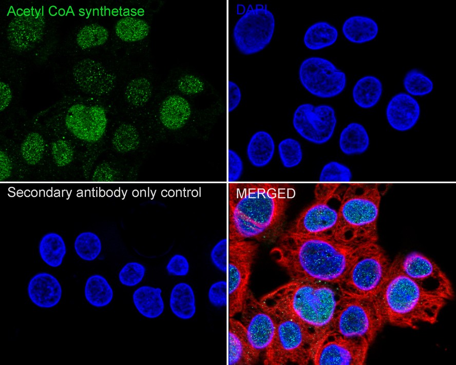
Immunocytochemistry analysis of HEPG2 cells labeling Acetyl CoA synthetase with Rabbit anti-Acetyl CoA synthetase antibody at 1/100 dilution. Cells were fixed in 100% precooled methanol for 5 minutes at room temperature, then blocked with 1% BSA in 10% negative goat serum for 1 hour at room temperature. Cells were then incubated with Rabbit anti-Acetyl CoA synthetase antibody at 1/100 dilution in 1% BSA in PBST overnight at 4 ℃. Goat Anti-Rabbit IgG H&L (488) was used as the secondary antibody at 1/1,000 dilution. PBS instead of the primary antibody was used as the secondary antibody only control. Nuclear DNA was labelled in blue with DAPI. Beta tubulin (red) was stained at 1/100 dilution overnight at +4℃. Goat Anti-Mouse IgG H&L (594) was used as the secondary antibody at 1/1,000 dilution.| Product Name | Acetyl CoA synthetase Recombinant Rabbit Monoclonal Antibody |
|---|---|
| Antibody Type | Primary Antibodies |
| Product description | ACSS2 (acyl-CoA synthetase short-chain family member 2), also known as ACAS2, ACS, ACSA or AceCS, is a 701 amino acid cytoplasmic protein that belongs to the ATP-dependent AMP-binding enzyme family. Existing as a monomer, ACSS2 functions to catalyze the ATP-dependent activation of acetate, a reaction that yields acetyl-CoA for use in energy generation and lipid synthesis. ACSS2 expression, which is highest in liver and kidney tissue, is regulated by the presence of unsaturated fatty acids and sterol regulatory element-binding proteins (SREBPs). Human ACSS2 exists as two alternatively spliced isoforms and shares 93% sequence identity with its mouse counterpart, suggesting a conserved role between species. |
| Immunogen | Recombinant protein within Human Acetyl CoA synthetase aa 101-145 / 701. |
| Clonality | Monoclonal |
|---|---|
| Isotype | IgG |
| Host Species | Recombinant rabbit |
| Tested Applications | ICC/IFWB |
| WB:1:1000-1:2000 ICC/IF:1:50-1:200 |
|
| Species Reactivity | HumanMouseRat |
| Concentration | 1mg/ml |
| Purification | Protein A |
| Alternative Names | ACAS2 antibody AceCS antibody Acetate CoA ligase antibody Acetate thiokinase antibody AcetateCoA ligase antibody Acetyl CoA synthetase antibody Acetyl Coenzyme A synthetase 2 (ADP forming) antibody Acetyl coenzyme A synthetase cytoplasmic antibody Acetyl-CoA synthetase antibody Acetyl-coenzyme A synthetase antibody ACS antibody ACSA antibody ACSA_HUMAN antibody ACSS2 antibody Acyl activating enzyme antibody Acyl CoA synthetase short chain family member 2 antibody Acyl-activating enzyme antibody Acyl-CoA synthetase short-chain family member 2 antibody Cytoplasmic acetyl coenzyme A synthetase antibody cytoplasmic antibody MYH7B antibody |
|---|---|
| Molecular Weight(MW) | 79 kDa |
| Cellular Localization | Cytoplasm, Nucleus. |
WB
Western blot analysis of Acetyl CoA synthetase on different lysates with Rabbit anti-Acetyl CoA synthetase antibody at 1/1,000 dilution. Lane 1: HepG2 cell lysate, Lane 2: L6 cell lysate, Lane 3: COS-1 cell lysate, Lysates/proteins at 20 µg/Lane. Exposure time: 3 minutes; 4-20% SDS-PAGE gel. Proteins were transferred to a PVDF membrane and blocked with 5% NFDM/TBST for 1 hour at room temperature. The primary antibody at 1/1,000 dilution was used in 5% NFDM/TBST at 4℃ overnight. Goat Anti-Rabbit IgG - HRP Secondary Antibody at 1/50,000 dilution was used for 1 hour at room temperature.
WB
Western blot analysis of Acetyl CoA synthetase on different lysates with Rabbit anti-Acetyl CoA synthetase antibody at 1/2,000 dilution. Lane 1: A549-si NT cell lysate, Lane 2: A549-si Acetyl CoA synthetase cell lysate, Lysates/proteins at 10 µg/Lane. Exposure time: 3 minutes; 4-20% SDS-PAGE gel. Proteins were transferred to a PVDF membrane and blocked with 5% NFDM/TBST for 1 hour at room temperature. The primary antibody at 1/2,000 dilution was used in 5% NFDM/TBST at 4℃ overnight. Goat Anti-Rabbit IgG - HRP Secondary Antibody at 1/50,000 dilution was used for 1 hour at room temperature.
IF
Immunocytochemistry analysis of HEPG2 cells labeling Acetyl CoA synthetase with Rabbit anti-Acetyl CoA synthetase antibody at 1/100 dilution. Cells were fixed in 100% precooled methanol for 5 minutes at room temperature, then blocked with 1% BSA in 10% negative goat serum for 1 hour at room temperature. Cells were then incubated with Rabbit anti-Acetyl CoA synthetase antibody at 1/100 dilution in 1% BSA in PBST overnight at 4 ℃. Goat Anti-Rabbit IgG H&L (488) was used as the secondary antibody at 1/1,000 dilution. PBS instead of the primary antibody was used as the secondary antibody only control. Nuclear DNA was labelled in blue with DAPI. Beta tubulin (red) was stained at 1/100 dilution overnight at +4℃. Goat Anti-Mouse IgG H&L (594) was used as the secondary antibody at 1/1,000 dilution.| Application Notes | WB:1:1000-1:2000 ICC/IF:1:50-1:200 |
|---|
| Form | Liquid |
|---|---|
| Storage Instructions | Store at +4℃ after thawing. Aliquot store at -20℃ or -80℃. Avoid repeated freeze / thaw cycles. |
| Storage Buffer | 1*TBS (pH7.4), 1%BSA, 40%Glycerol. Preservative: 0.05% Sodium Azide. |


