Application
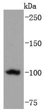
Fig1: Western blot analysis of TERT on Hela cells lysates using anti-TERT antibody at 1/1,000 dilution.Application
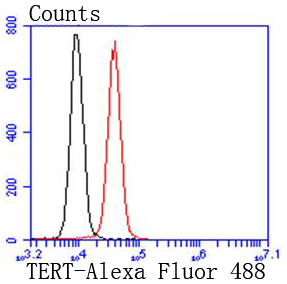
Fig2: Flow cytometric analysis of Hela cells with TERT antibody at 1/50 dilution (red) compared with an unlabelled control (cells without incubation with primary antibody; black). Alexa Fluor 488-conjugated goat anti rabbit IgG was used as the secondary antibody| Product Name | Telomerase reverse transcriptase [JF0586] |
|---|---|
| Antibody Type | Primary Antibodies |
| Antigen Alias | CMM9 antibody DKCA2 antibody DKCB4 antibody EST2 antibody HEST2 antibody htert antibody hTRT antibody PFBMFT1 antibody TCS1 antibody Telomerase associated protein 2 antibody Telomerase catalytic subunit antibody Telomerase reverse transcriptase antibody Telomerase-associated protein 2 antibody Telomere Reverse Transcriptase antibody TERT antibody TERT_HUMAN antibody TP2 antibody TRT antibody |
| Product description | Telomerase is an RNA-dependent DNA polymerase that catalyzes the addition of telomeric repeat sequences to chromosome ends. In most human somatic cells, telomerase activity is undetectable, and telomeres shorten with successive cell divisions. However, telomerase activity is detectable in immortal cells and in many human tumors. Two candidate mammalian telomerase proteins have been cloned. Human TP1 (for telomerase-associated protein 1), also designated TLP1 in rat (for telomerase protein component 1), is homologous to the Tetrahymena p80 telomerase protein and has been shown to interact with mammalian telomerase RNA. Human TERT (for telomerase reverse transcriptase), also designated hEST2 (for ever shorter telomeres), is homologous to the p123 telomerase protein from Euplotes and to the yeast Est2 protein. Expression of TERT mRNA has been shown to correlate with telomerase activity in various cell lines. |
| Immunogen | recombinant protein |
| Clonality | Monoclonal |
|---|---|
| Isotype | IgG |
| Host Species | Recombinant rabbit |
| Tested Applications | WBFC |
| WB:1:1,000-1:2,000 FC:1:50-1:100 |
|
| Species Reactivity | Human |
| Concentration | 1mg/ml |
| Alternative Names | CMM9 antibody DKCA2 antibody DKCB4 antibody EST2 antibody HEST2 antibody htert antibody hTRT antibody PFBMFT1 antibody TCS1 antibody Telomerase associated protein 2 antibody Telomerase catalytic subunit antibody Telomerase reverse transcriptase antibody Telomerase-associated protein 2 antibody Telomere Reverse Transcriptase antibody TERT antibody TERT_HUMAN antibody TP2 antibody TRT antibody |
|---|---|
| Molecular Weight(MW) | 127/120/90/89 kDa |
| Cellular Localization | Nucleus, Chromosome, Cytoplasm. |
| SwissProt ID | O14746 |
|---|
Application
Fig1: Western blot analysis of TERT on Hela cells lysates using anti-TERT antibody at 1/1,000 dilution.
Application
Fig2: Flow cytometric analysis of Hela cells with TERT antibody at 1/50 dilution (red) compared with an unlabelled control (cells without incubation with primary antibody; black). Alexa Fluor 488-conjugated goat anti rabbit IgG was used as the secondary antibody| Positive Control | Hela. |
|---|---|
| Application Notes | WB:1:1,000-1:2,000 FC:1:50-1:100 |
| Form | Liquid |
|---|---|
| Storage Instructions | Store at +4℃ after thawing. Aliquot store at -20℃ or -80℃. Avoid repeated freeze / thaw cycles. |
| Storage Buffer | 1*TBS (pH7.4), 1%BSA, 40%Glycerol. Preservative: 0.05% Sodium Azide. |


