Application
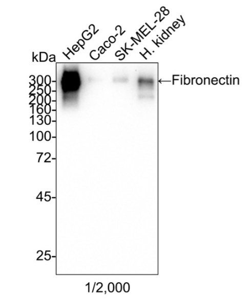
Fig1: Western blot analysis of Fibronectin on different lysates with Rabbit anti-Fibronectin antibody at 1/2,000 dilution and competitor's antibody at 1/2,000 dilution. Lane 1: HepG2 cell lysate, Lane 2: Caco-2 cell lysate, Lane 3: SK-MEL-28 cell lysate, Lane 4: Human kidney tissue lysate, Lysates/proteins at 20 µg/Lane. Exposure time: 3 minutes 20 seconds; 4-20% SDS-PAGE gel. Proteins were transferred to a PVDF membrane and blocked with 5% NFDM/TBST for 1 hour at room temperature. The primary antibody at 1/2,000 dilution and competitor's antibody at 1/2,000 dilution were used in 5% NFDM/TBST at 4℃ overnight. Goat Anti-Rabbit IgG - HRP Secondary Antibody at 1/50,000 dilution was used for 1 hour at room temperature.Application
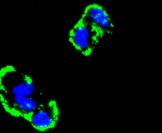
Fig2: ICC staining Fibronectin in Hela cells (green). The nuclear counter stain is DAPI (blue). Cells were fixed in paraformaldehyde, permeabilised with 0.25% Triton X100/PBS.Application
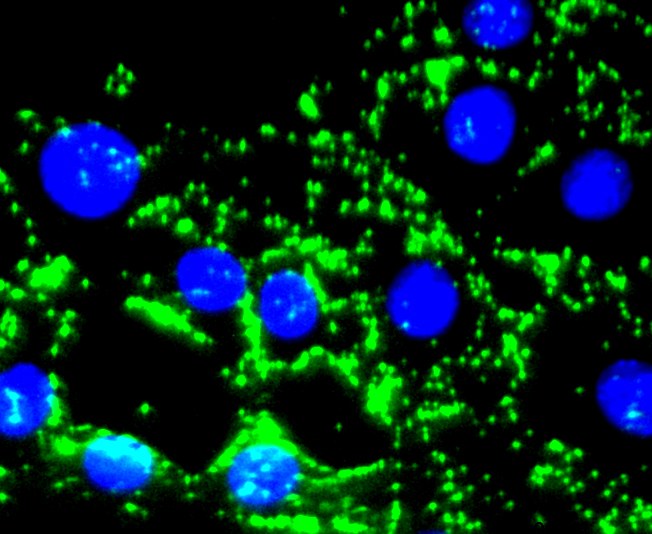
Fig3: ICC staining Fibronectin in NIH/3T3 cells (green). The nuclear counter stain is DAPI (blue). Cells were fixed in paraformaldehyde, permeabilised with 0.25% Triton X100/PBS.Application
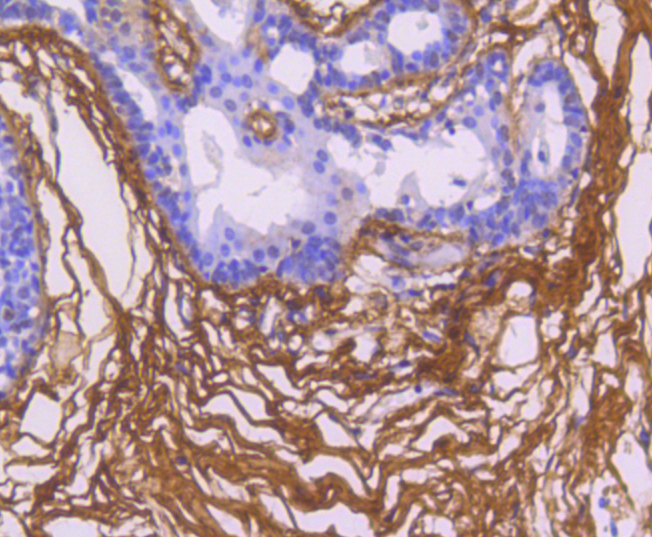
Fig4: Immunohistochemical analysis of paraffin-embedded human breast carcinoma tissue using anti-Fibronectin antibody. Counter stained with hematoxylin.Application
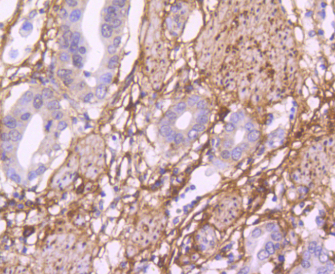
Fig5: Immunohistochemical analysis of paraffin-embedded human gastric carcinoma tissue using anti-Fibronectin antibody. Counter stained with hematoxylin.| Product Name | Fibronectin Recombinant Rabbit Monoclonal Antibody |
|---|---|
| Antibody Type | Primary Antibodies |
| Product description | Fibronectin is an extracellular matrix glycoprotein present on most cell surfaces, in extracellular fluids and in plasma. A high molecular weight heterodimeric protein, it was originally discovered as a protein missing from the surfaces of virus-transformed cells, and it has been shown to be involved in various functions including cell adhesion, cell motility and wound healing. Alternative splicing and glycosylation give rise to several different forms of Fibronectin, some of which exhibit restricted tissue distribution or association with malignancies. It has been shown that Myofibroblast phenotype formation correlates with the occurrence of glycosylated Fibronectin and Fibronectin splice variants in Dupuytren's disease. |
| Immunogen | Recombinant full length protein of Human Fibronectin aa 1-2477 / 2477. |
| Clonality | monoclonal |
|---|---|
| Isotype | IgG |
| Host Species | Recombinant rabbit |
| Tested Applications | ICC/IFIHCWB |
| WB:1:1,000 ICC:1:100-1:500 IHC:1:50-1:200 |
|
| Species Reactivity | HumanMouse |
| Concentration | 1mg/ml |
| Purification | Protein A |
| Alternative Names | CIG antibody Cold insoluble globulin antibody Cold-insoluble globulin antibody DKFZp686F10164 antibody DKFZp686H0342 antibody DKFZp686I1370 antibody DKFZp686O13149 antibody ED B antibody Fibronectin 1 antibody FINC antibody FINC_HUMAN antibody FN antibody FN1 antibody FNZ antibody GFND antibody GFND2 antibody LETS antibody Migration stimulating factor antibody MSF antibody Ugl-Y3 antibody |
|---|---|
| Molecular Weight(MW) | 22 kDa |
| Cellular Localization | Secreted. |
| SwissProt ID | P02751 |
|---|
Application
Fig1: Western blot analysis of Fibronectin on different lysates with Rabbit anti-Fibronectin antibody at 1/2,000 dilution and competitor's antibody at 1/2,000 dilution. Lane 1: HepG2 cell lysate, Lane 2: Caco-2 cell lysate, Lane 3: SK-MEL-28 cell lysate, Lane 4: Human kidney tissue lysate, Lysates/proteins at 20 µg/Lane. Exposure time: 3 minutes 20 seconds; 4-20% SDS-PAGE gel. Proteins were transferred to a PVDF membrane and blocked with 5% NFDM/TBST for 1 hour at room temperature. The primary antibody at 1/2,000 dilution and competitor's antibody at 1/2,000 dilution were used in 5% NFDM/TBST at 4℃ overnight. Goat Anti-Rabbit IgG - HRP Secondary Antibody at 1/50,000 dilution was used for 1 hour at room temperature.
Application
Fig2: ICC staining Fibronectin in Hela cells (green). The nuclear counter stain is DAPI (blue). Cells were fixed in paraformaldehyde, permeabilised with 0.25% Triton X100/PBS.
Application
Fig3: ICC staining Fibronectin in NIH/3T3 cells (green). The nuclear counter stain is DAPI (blue). Cells were fixed in paraformaldehyde, permeabilised with 0.25% Triton X100/PBS.
Application
Fig4: Immunohistochemical analysis of paraffin-embedded human breast carcinoma tissue using anti-Fibronectin antibody. Counter stained with hematoxylin.
Application
Fig5: Immunohistochemical analysis of paraffin-embedded human gastric carcinoma tissue using anti-Fibronectin antibody. Counter stained with hematoxylin.| Application Notes | WB:1:1,000 ICC:1:100-1:500 IHC:1:50-1:200 |
|---|
| Form | Liquid |
|---|---|
| Storage Instructions | Store at +4℃ after thawing. Aliquot store at -20℃ or -80℃. Avoid repeated freeze / thaw cycles. |
| Storage Buffer | 1*TBS (pH7.4), 1%BSA, 40%Glycerol. Preservative: 0.05% Sodium Azide. |


