Application
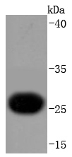
Fig1: Western blot analysis of Troponin I on human heart lysates using anti-Troponin I antibody at 1/1,000 dilution.Application
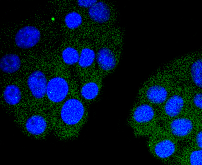
Fig2: ICC staining Troponin I in HepG2 cells (green). The nuclear counter stain is DAPI (blue). Cells were fixed in paraformaldehyde, permeabilised with 0.25% Triton X100/PBS.Application
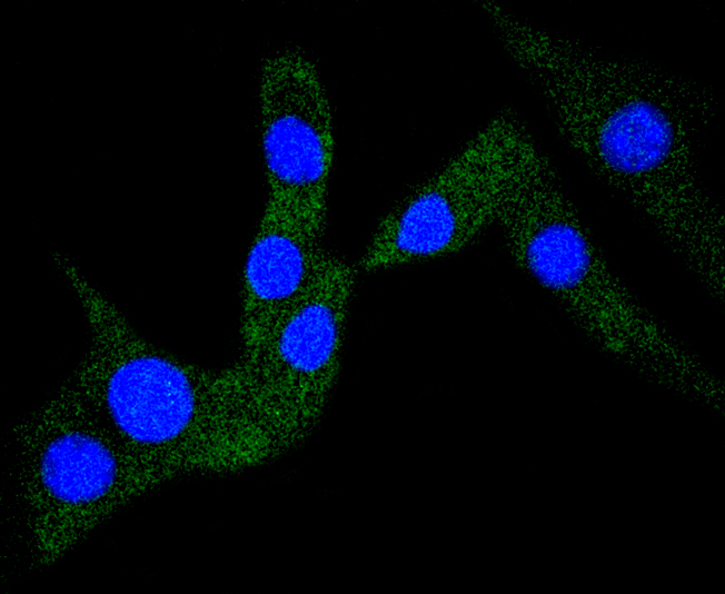
Fig3: ICC staining Troponin I in NIH/3T3 cells (green). The nuclear counter stain is DAPI (blue). Cells were fixed in paraformaldehyde, permeabilised with 0.25% Triton X100/PBS.Application
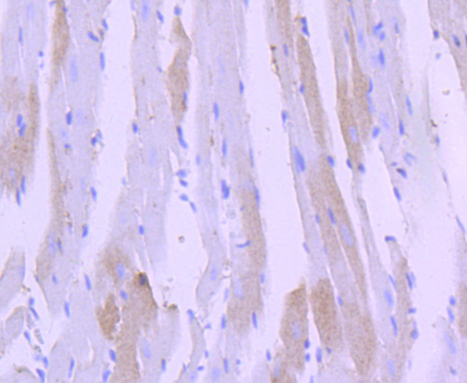
Fig4: Immunohistochemical analysis of paraffin-embedded human heart tissue using anti-Troponin I antibody. Counter stained with hematoxylin.| Product Name | Cardiac Troponin I [JF0550] |
|---|---|
| Antibody Type | Primary Antibodies |
| Antigen Alias | cardiac muscle antibody Cardiac troponin I antibody cardiomyopathy, dilated 2A (autosomal recessive) antibody Cardiomyopathy, familial hypertrophic, 7, included antibody CMD1FF antibody CMD2A antibody CMH7 antibody cTnI antibody Familial hypertrophic cardiomyopathy 7 antibody MGC116817 antibody RCM1 antibody Tn1 antibody Tni antibody TNN I3 antibody TNNC 1 antibody TNNC1 antibody TNNI3 antibody TNNI3_HUMAN antibody Troponin I antibody Troponin I cardiac antibody Troponin I cardiac muscle antibody Troponin I cardiac muscle isoform antibody Troponin I type 3 cardiac antibody troponin I, cardiac 3 antibody TroponinI antibody Ttroponin I type 3 (cardiac) antibody |
| Product description | Actin is a highly conserved protein that is expressed in all eukaryotic cells. Actin filaments can form both stable and labile structures and are crucial components of microvilli and the contractile apparatus of muscle cells. Myosin is a hexamer composed of two heavy chains (MHC) and four light chains (MLC); it interacts with Actin to generate the force for diverse cellular movements, including cytokinesis, phagocytosis and muscle contraction. Troponin facilitates the interaction between Actin and Myosin by binding to calcium. Troponin comprises at least two subunits, which are divergent in cardiac muscle, fast skeletal muscle and slow skeletal muscle. Structures of skeletal muscle troponin are composed of Troponin C (the sensor), Troponin I (the regulator) and Troponin T (the link to the muscle thin filament). Troponin C is dumbbell-shaped and has a hydrophobic pocket that increases the contractile force of muscle fibers. Troponin C has two isoforms: fast and slow. Fast Troponin C has two calcium binding sites while slow/cardiac Troponin C has a single calcium binding site. |
| Immunogen | recombinant protein |
| Clonality | Monoclonal |
|---|---|
| Isotype | IgG |
| Host Species | Recombinant rabbit |
| Tested Applications | WBICC/IFIHCIP |
| WB:1:1,000-1:5,000 ICC:1:50-1:200 IHC:1:50-1:200 |
|
| Species Reactivity | HumanMouse |
| Concentration | 1mg/ml |
| Alternative Names | cardiac muscle antibody
Cardiac troponin I antibody cardiomyopathy dilated 2A (autosomal recessive) antibody Cardiomyopathy familial hypertrophic 7 included antibody CMD1FF antibody CMD2A antibody CMH7 antibody cTnI antibody Familial hypertrophic cardiomyopathy 7 antibody MGC116817 antibody RCM1 antibody Tn1 antibody Tni antibody TNN I3 antibody TNNC 1 antibody TNNC1 antibody TNNI3 antibody TNNI3_HUMAN antibody Troponin I antibody Troponin I cardiac antibody Troponin I cardiac muscle antibody Troponin I cardiac muscle isoform antibody Troponin I type 3 cardiac antibody troponin I cardiac 3 antibody TroponinI antibody Ttroponin I type 3 (cardiac) antibody |
|---|---|
| Molecular Weight(MW) | 24 kDa |
| Cellular Localization | Cytosol. |
Application
Fig1: Western blot analysis of Troponin I on human heart lysates using anti-Troponin I antibody at 1/1,000 dilution.
Application
Fig2: ICC staining Troponin I in HepG2 cells (green). The nuclear counter stain is DAPI (blue). Cells were fixed in paraformaldehyde, permeabilised with 0.25% Triton X100/PBS.
Application
Fig3: ICC staining Troponin I in NIH/3T3 cells (green). The nuclear counter stain is DAPI (blue). Cells were fixed in paraformaldehyde, permeabilised with 0.25% Triton X100/PBS.
Application
Fig4: Immunohistochemical analysis of paraffin-embedded human heart tissue using anti-Troponin I antibody. Counter stained with hematoxylin.| Positive Control | NIH/3T3, HepG2, human heart tissue. |
|---|---|
| Application Notes | WB:1:1,000-1:5,000 ICC:1:50-1:200 IHC:1:50-1:200 |
| Form | Liquid |
|---|---|
| Storage Instructions | Store at +4℃ after thawing. Aliquot store at -20℃ or -80℃. Avoid repeated freeze / thaw cycles. |
| Storage Buffer | 1*TBS (pH7.4), 1%BSA, 40%Glycerol. Preservative: 0.05% Sodium Azide. |


