WB
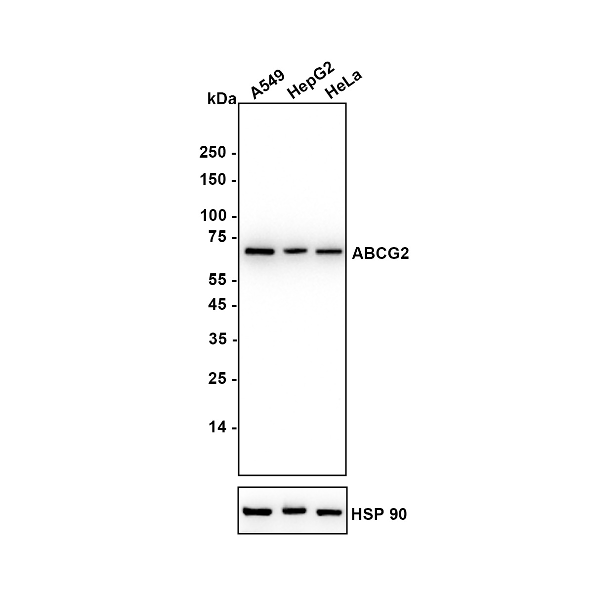
Western blot analysis of ABCG2 on different lysates with Rabbit anti-ABCG2 antibody at 1/2,000 dilution. Lane 1: A549 (Human lung adenocarcinoma cell), Lane 2: Hep G2 (Human hepatocellular carcinoma cell), Lane 3: HeLa (Human cervical adenocarcinoma cell), Lysates/proteins at 20 µg/Lane. Exposure time: 12 seconds; Blocking: 5% NFDM/TBST, 1 hour at room temperature Primary antibody: 1/2,000 in primary antibody dilution buffer, overnight at 4 ℃ Secondary antibody: Goat anti-Rabbit IgG-HRP, 1/50,000 in 5% NFDM/TBST, 1 hour at room temperature.IHC
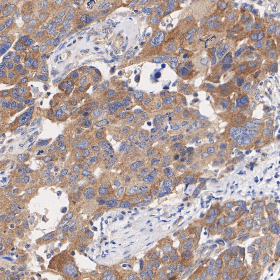
Immunohistochemical analysis of paraffin-embedded human ovarian cancer tissue with Rabbit anti-ABCG2 antibody at 1/200 dilution. The section was pre-treated using heat mediated antigen retrieval with Tris-EDTA buffer (pH 9.0) for 20 minutes. The tissues were blocked in 1% BSA for 20 minutes at room temperature, washed with ddH2O and PBS, and then probed with the primary antibody at 1/200 dilution for 1 hour at room temperature. The detection was performed using an HRP conjugated compact polymer system. DAB was used as the chromogen. Tissues were counterstained with hematoxylin and mounted with DPX.IHC
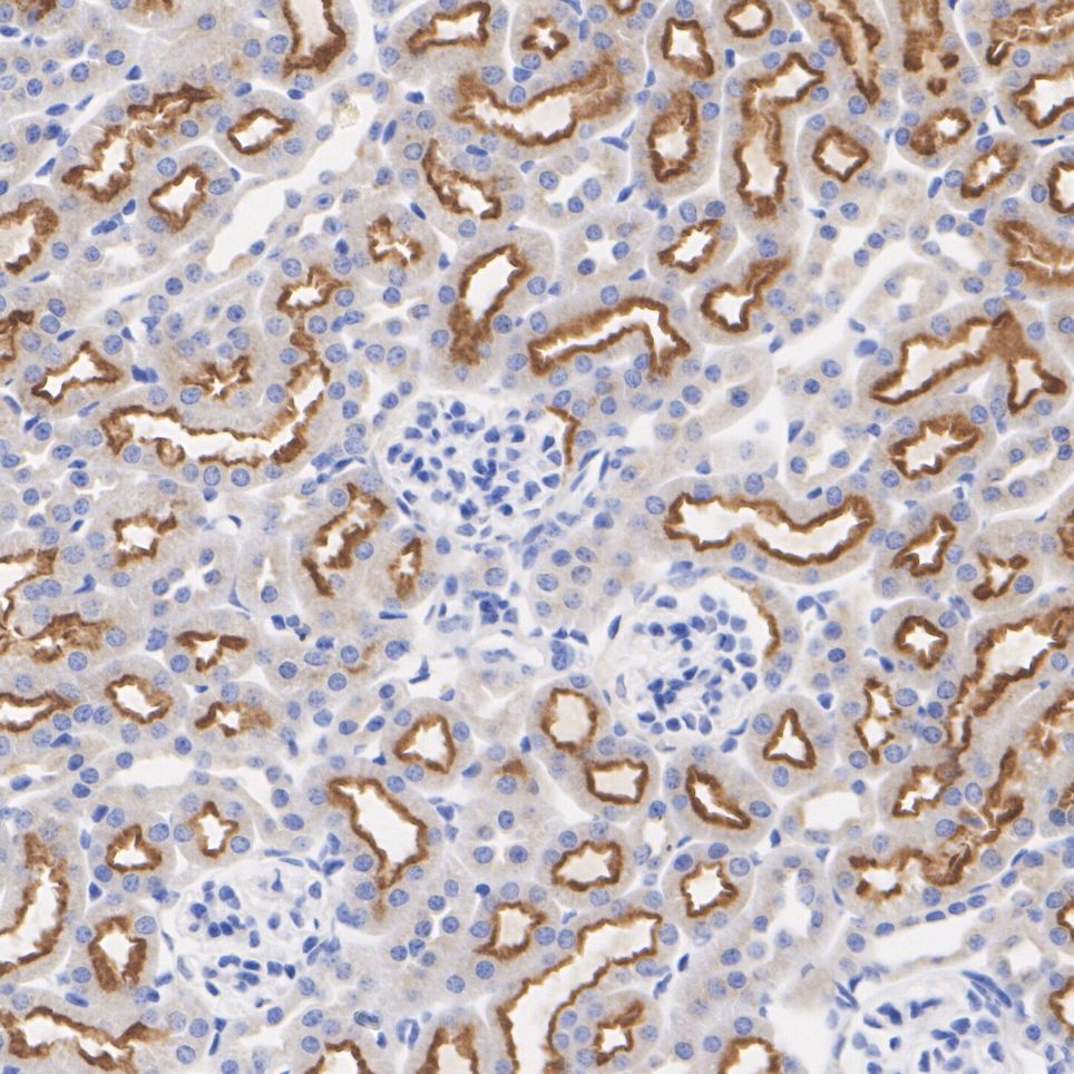
Immunohistochemical analysis of paraffin-embedded mouse kidney tissue with Rabbit anti-ABCG2 antibody at 1/1,000 dilution. The section was pre-treated using heat mediated antigen retrieval with Tris-EDTA buffer (pH 9.0) for 20 minutes. The tissues were blocked in 1% BSA for 20 minutes at room temperature, washed with ddH2O and PBS, and then probed with the primary antibody at 1/1,000 dilution for 1 hour at room temperature. The detection was performed using an HRP conjugated compact polymer system. DAB was used as the chromogen. Tissues were counterstained with hematoxylin and mounted with DPX.| Product Name | ABCG2 Recombinant Rabbit Monoclonal Antibody |
|---|---|
| Antibody Type | Primary Antibodies |
| Product description | ATP-binding cassette (ABC) transporters are an evolutionarily conserved family of proteins that catalyze the transport of molecules across extracellular and intracellular membranes through the energy of ATP hydrolysis. The ABC half-transporter, ABCG2, is also known as placenta-specific ABC transporter and breast cancer resistance protein (BCRP1). ABCG2 confers resistance for a variety of chemotherapeutic agents, including anthracyclines, mitoxantrone, bisantrene and topotecan. Under normal conditions, ABCG2 may serve a protective function by removing toxins from the cell, and plays an important role in regulating stem cell differentiation. ABCG2 is responsible for the side population (SP) phenotype and is widely expressed in a large variety of stem cells, making it an important stem cell marker. ABCG2 may have N-linked glycosylation and may dimerize in vivo. ABCG2 is abundantly expressed in placenta, liver, intestine and stem cells. |
| Immunogen | Synthetic peptide within human ABCG2 aa 140-180. |
| Clonality | monoclonal |
|---|---|
| Isotype | IgG |
| Host Species | Recombinant rabbit |
| Tested Applications | IHCWB |
| WB:1:500-1:2000 IHC:1:50-1:200 |
|
| Species Reactivity | HumanMouse |
| Concentration | 1mg/ml |
| Purification | Protein A |
| Alternative Names | ABC transporter antibody ABC15 antibody ABCG 2 antibody ABCG2 antibody ABCG2_HUMAN antibody ABCP antibody ATP binding cassette sub family G (WHITE) member 2 antibody ATP binding cassette transporter G2 antibody ATP-binding cassette sub-family G member 2 antibody BCRP antibody BCRP1 antibody BMDP antibody Breast cancer resistance protein antibody CD338 antibody CDw338 antibody CDw338 antigen antibody EST157481 antibody GOUT1 antibody MGC102821 antibody Mitoxantrone resistance associated protein antibody Mitoxantrone resistance-associated protein antibody MRX antibody Multi drug resistance efflux transport ATP binding cassette sub family G (WHITE) member 2 antibody MXR antibody MXR1 antibody Placenta specific ATP binding cassette transporter antibody Placenta specific MDR protein antibody Placenta-specific ATP-binding cassette transporter antibody UAQTL1 antibody |
|---|---|
| Molecular Weight(MW) | 72 kDa |
| Cellular Localization | Cell membrane, Mitochondrion membrane. |
WB
Western blot analysis of ABCG2 on different lysates with Rabbit anti-ABCG2 antibody at 1/2,000 dilution. Lane 1: A549 (Human lung adenocarcinoma cell), Lane 2: Hep G2 (Human hepatocellular carcinoma cell), Lane 3: HeLa (Human cervical adenocarcinoma cell), Lysates/proteins at 20 µg/Lane. Exposure time: 12 seconds; Blocking: 5% NFDM/TBST, 1 hour at room temperature Primary antibody: 1/2,000 in primary antibody dilution buffer, overnight at 4 ℃ Secondary antibody: Goat anti-Rabbit IgG-HRP, 1/50,000 in 5% NFDM/TBST, 1 hour at room temperature.
IHC
Immunohistochemical analysis of paraffin-embedded human ovarian cancer tissue with Rabbit anti-ABCG2 antibody at 1/200 dilution. The section was pre-treated using heat mediated antigen retrieval with Tris-EDTA buffer (pH 9.0) for 20 minutes. The tissues were blocked in 1% BSA for 20 minutes at room temperature, washed with ddH2O and PBS, and then probed with the primary antibody at 1/200 dilution for 1 hour at room temperature. The detection was performed using an HRP conjugated compact polymer system. DAB was used as the chromogen. Tissues were counterstained with hematoxylin and mounted with DPX.
IHC
Immunohistochemical analysis of paraffin-embedded mouse kidney tissue with Rabbit anti-ABCG2 antibody at 1/1,000 dilution. The section was pre-treated using heat mediated antigen retrieval with Tris-EDTA buffer (pH 9.0) for 20 minutes. The tissues were blocked in 1% BSA for 20 minutes at room temperature, washed with ddH2O and PBS, and then probed with the primary antibody at 1/1,000 dilution for 1 hour at room temperature. The detection was performed using an HRP conjugated compact polymer system. DAB was used as the chromogen. Tissues were counterstained with hematoxylin and mounted with DPX.| Application Notes | WB:1:500-1:2000 IHC:1:50-1:200 |
|---|
| Form | Liquid |
|---|---|
| Storage Instructions | Store at +4℃ after thawing. Aliquot store at -20℃ or -80℃. Avoid repeated freeze / thaw cycles. |
| Storage Buffer | 1*TBS (pH7.4), 1%BSA, 40%Glycerol. Preservative: 0.05% Sodium Azide. |


