WB
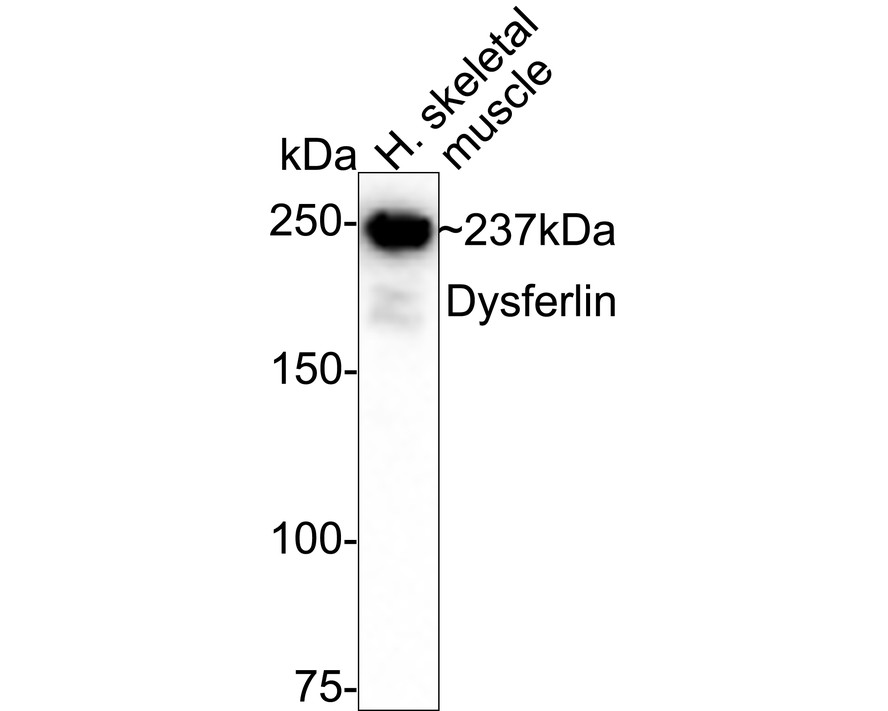
Western blot analysis of Dysferlin on human skeletal muscle tissue lysates with Rabbit anti-Dysferlin antibody at 1/1,000 dilution. Lysates/proteins at 20 µg/Lane. Exposure time: 3 minutes; 6% SDS-PAGE gel. Proteins were transferred to a PVDF membrane and blocked with 5% NFDM/TBST for 1 hour at room temperature. The primary antibody at 1/1,000 dilution was used in 5% NFDM/TBST at room temperature for 2 hours. Goat Anti-Rabbit IgG - HRP Secondary Antibody at 1/50,000 dilution was used for 1 hour at room temperature.IHC
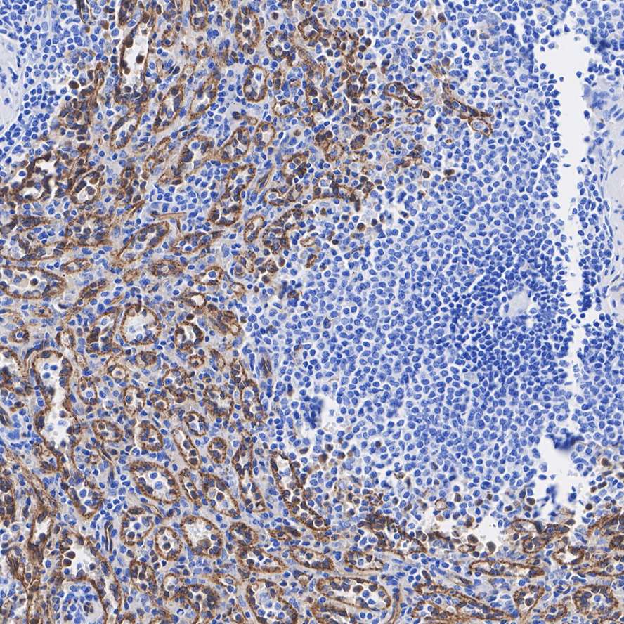
Immunohistochemical analysis of paraffin-embedded human spleen tissue with Rabbit anti-Dysferlin antibody at 1/200 dilution. The section was pre-treated using heat mediated antigen retrieval with Tris-EDTA buffer (pH 9.0) for 20 minutes. The tissues were blocked in 1% BSA for 20 minutes at room temperature, washed with ddH2O and PBS, and then probed with the primary antibody at 1/200 dilution for 1 hour at room temperature. The detection was performed using an HRP conjugated compact polymer system. DAB was used as the chromogen. Tissues were counterstained with hematoxylin and mounted with DPX.IHC
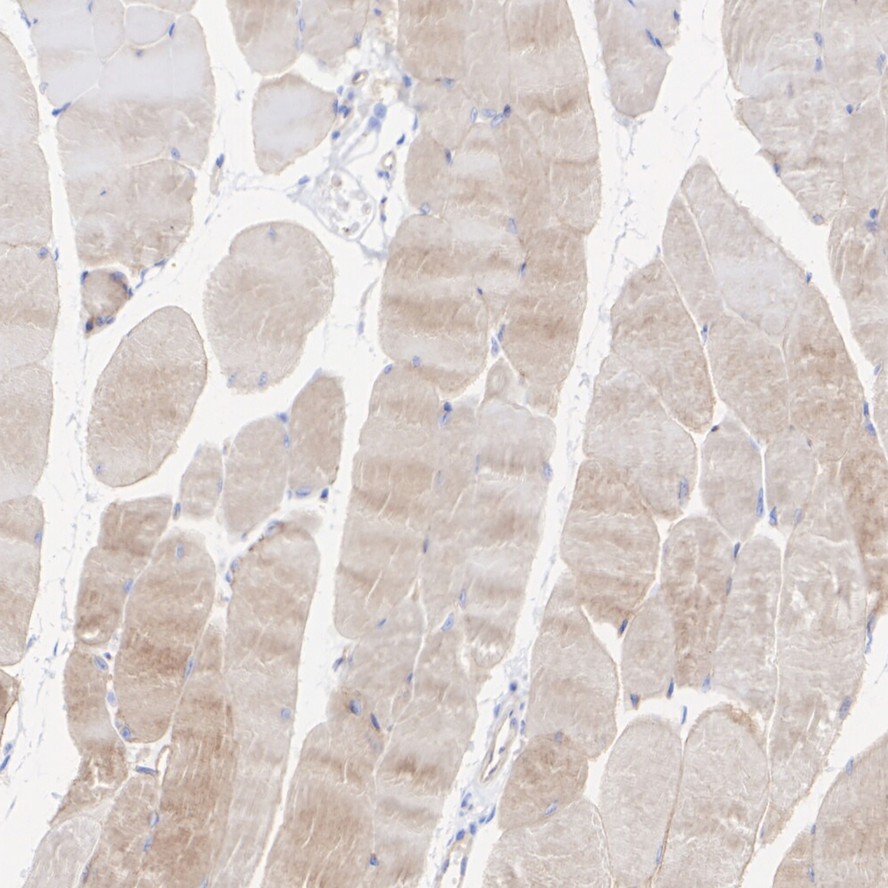
Immunohistochemical analysis of paraffin-embedded mouse skeletal muscle tissue with Rabbit anti-Dysferlin antibody at 1/200 dilution. The section was pre-treated using heat mediated antigen retrieval with Tris-EDTA buffer (pH 9.0) for 20 minutes. The tissues were blocked in 1% BSA for 20 minutes at room temperature, washed with ddH2O and PBS, and then probed with the primary antibody at 1/200 dilution for 1 hour at room temperature. The detection was performed using an HRP conjugated compact polymer system. DAB was used as the chromogen. Tissues were counterstained with hematoxylin and mounted with DPX.| Product Name | Dysferlin Recombinant Rabbit Monoclonal Antibody |
|---|---|
| Antibody Type | Primary Antibodies |
| Product description | Dysferlin is a muscle-specific protein that is essential for normal muscle function and development. Mutations in the human dysferlin gene, DYSF, which maps to chromosome 2p13.2, are associated with limb girdle muscular dystrophy-2B (LGMD-2B) and a related, adult-onset, distal dystrophy known as Miyoshi myopathy (MM). Dysferlin localizes to the muscle fiber membrane, but is absent in MM and LGMD-2B muscle. Dysferlin is detected in 5-6 week embryos, when limbs begin to form regional differentiation. Although it is not essential for initial myogenesis, dysferlin appears to be critical for sustained normal function in mature muscle. It has been suggested that the absence of dysferlin during development gives rise to the disease phenotype in adulthood. Identical mutations in the dysferlin gene can produce more than one myopathy phenotype, indicating that additional genes and/or other factors are also involved in the clinical phenotype. The DYSF gene has no homology to any other known mammalian gene, but the protein product is related to the spermatogenesis factor fer-1 of Caenorhabditis elegans. The name “dysferlin” combines the role of the gene in producing muscular dystrophy with its homology to C. elegans. |
| Immunogen | Synthetic peptide within Human Dysferlin aa 113-162 / 2080. |
| Clonality | Monoclonal |
|---|---|
| Isotype | IgG |
| Host Species | Recombinant rabbit |
| Tested Applications | IHCWB |
| WB:1:1000-1:5000 IHC:1:200 |
|
| Species Reactivity | HumanMouse |
| Concentration | 1mg/ml |
| Purification | Protein A |
| Alternative Names | DMAT antibody DYSF antibody DYSF_HUMAN antibody Dysferlin antibody Dysferlin limb girdle muscular dystrophy 2B (autosomal recessive) antibody Dysferlin limb girdle muscular dystrophy 2B antibody Dystrophy associated fer 1 like 1 antibody Dystrophy associated fer 1 like protein antibody Dystrophy associated fer1 like 1 antibody Dystrophy associated fer1 like protein antibody Dystrophy-associated fer-1-like protein antibody Fer 1 like protein 1 antibody Fer-1-like protein 1 antibody Fer1 like protein 1 antibody FER1L1 antibody FLJ00175 antibody FLJ90168 antibody LGMD 2B antibody LGMD2B antibody Limb girdle muscular dystrophy 2B (autosomal recessive) antibody Limb girdle muscular dystrophy 2B antibody Miyoshi myopathy antibody MM antibody MMD1 antibody |
|---|---|
| Molecular Weight(MW) | 237 kDa |
| Cellular Localization | Cell membrane, Cytoplasmic vesicle membrane. |
WB
Western blot analysis of Dysferlin on human skeletal muscle tissue lysates with Rabbit anti-Dysferlin antibody at 1/1,000 dilution. Lysates/proteins at 20 µg/Lane. Exposure time: 3 minutes; 6% SDS-PAGE gel. Proteins were transferred to a PVDF membrane and blocked with 5% NFDM/TBST for 1 hour at room temperature. The primary antibody at 1/1,000 dilution was used in 5% NFDM/TBST at room temperature for 2 hours. Goat Anti-Rabbit IgG - HRP Secondary Antibody at 1/50,000 dilution was used for 1 hour at room temperature.
IHC
Immunohistochemical analysis of paraffin-embedded human spleen tissue with Rabbit anti-Dysferlin antibody at 1/200 dilution. The section was pre-treated using heat mediated antigen retrieval with Tris-EDTA buffer (pH 9.0) for 20 minutes. The tissues were blocked in 1% BSA for 20 minutes at room temperature, washed with ddH2O and PBS, and then probed with the primary antibody at 1/200 dilution for 1 hour at room temperature. The detection was performed using an HRP conjugated compact polymer system. DAB was used as the chromogen. Tissues were counterstained with hematoxylin and mounted with DPX.
IHC
Immunohistochemical analysis of paraffin-embedded mouse skeletal muscle tissue with Rabbit anti-Dysferlin antibody at 1/200 dilution. The section was pre-treated using heat mediated antigen retrieval with Tris-EDTA buffer (pH 9.0) for 20 minutes. The tissues were blocked in 1% BSA for 20 minutes at room temperature, washed with ddH2O and PBS, and then probed with the primary antibody at 1/200 dilution for 1 hour at room temperature. The detection was performed using an HRP conjugated compact polymer system. DAB was used as the chromogen. Tissues were counterstained with hematoxylin and mounted with DPX.| Application Notes | WB:1:1000-1:5000 IHC:1:200 |
|---|
| Form | Liquid |
|---|---|
| Storage Instructions | Store at +4℃ after thawing. Aliquot store at -20℃ or -80℃. Avoid repeated freeze / thaw cycles. |
| Storage Buffer | 1*TBS (pH7.4), 1%BSA, 40%Glycerol. Preservative: 0.05% Sodium Azide. |


