Application
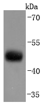
Fig1: Western blot analysis of CNPase on mouse brain lysates using anti-CNPase antibody at 1/1,000 dilution.Application
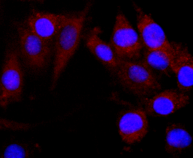
Fig2: ICC staining CNPase in SHG-44 cells (green). The nuclear counter stain is DAPI (blue). Cells were fixed in paraformaldehyde, permeabilised with 0.25% Triton X100/PBS.Application
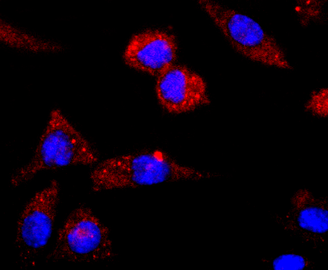
Fig3: ICC staining CNPase in SH-SY-5Y cells (green). The nuclear counter stain is DAPI (blue). Cells were fixed in paraformaldehyde, permeabilised with 0.25% Triton X100/PBS.Application
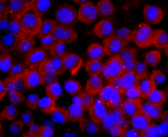
Fig4: ICC staining CNPase in N2A cells (green). Cells were fixed in paraformaldehyde, permeabilised with 0.25% Triton X100/PBS.Application
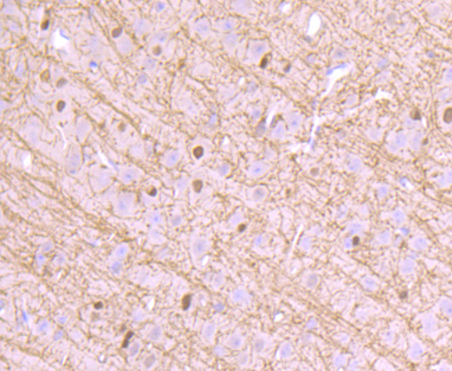
Fig5: Immunohistochemical analysis of paraffin-embedded mouse brain tissue using anti-CNPase antibody. Counter stained with hematoxylin.Application
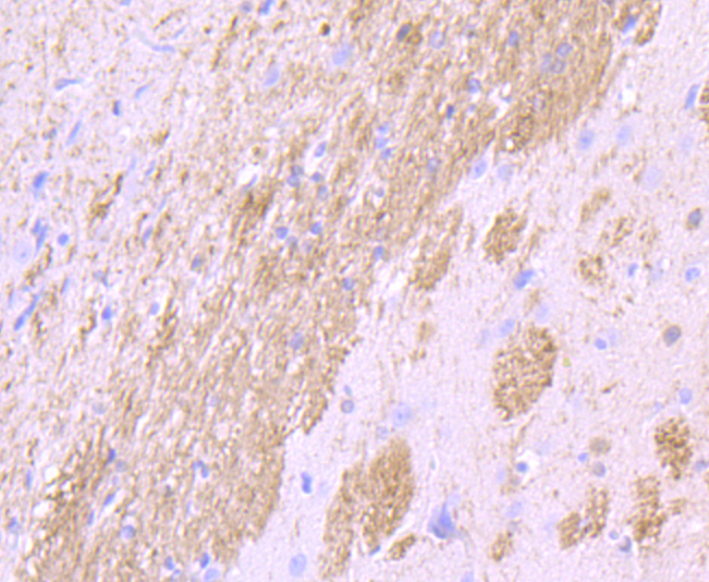
Fig6: Immunohistochemical analysis of paraffin-embedded rat brain tissue using anti-CNPase antibody. Counter stained with hematoxylin.Application
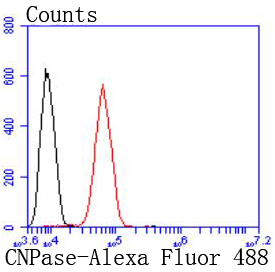
Fig7: Flow cytometric analysis of SH-SY-5Y cells with CNPase antibody at 1/50 dilution (blue) compared with an unlabelled control (cells without incubation with primary antibody; red). Alexa Fluor 488-conjugated goat anti-rabbit IgG was used as the secondary antibody.| Product Name | CNPase [JF10-25] |
|---|---|
| Antibody Type | Primary Antibodies |
| Antigen Alias | 2'' antibody 2'3' cyclic nucleotide 3' phosphodiesterase antibody 3''-cyclic-nucleotide 3''-phosphodiesterase antibody CN37_HUMAN antibody CNP 1 antibody CNP antibody CNP1 antibody CNPase antibody |
| Product description | 2',3'-cyclic nucleotide-3'-phosphodiesterase (CNPase) is a membrane-bound enzyme that can link tubulin to membranes and may regulate cytoplasmic microtubule distribution. CNPase acts as a microtubule-associated protein by promoting microtubule assembly; this activity resides in the C-terminus of the enzyme. CNPase is firmly associated with tubulin from brain tissue and thyroid cells and can be found at high concentrations in central nervous system myelin and in the outer segments of photoreceptors in the retina. Acute lead intoxication leads to disturbances in CNPase activity and the morphology of myelin. |
| Immunogen | recombinant protein |
| Clonality | Monoclonal |
|---|---|
| Isotype | IgG |
| Host Species | Recombinant rabbit |
| Tested Applications | WBICCIHCFC |
| WB:1:1,000-1:2,000 ICC:1:50-1:200 IHC:1:50-1:200 FC:1:50-1:100 |
|
| Species Reactivity | HumanMouseRat |
| Concentration | 1mg/ml |
| Alternative Names | 2'' antibody 2'3' cyclic nucleotide 3' phosphodiesterase antibody 3''-cyclic-nucleotide 3''-phosphodiesterase antibody CN37_HUMAN antibody CNP 1 antibody CNP antibody CNP1 antibody CNPase antibody |
|---|---|
| Molecular Weight(MW) | 48 kDa |
| Cellular Localization | Membrane, Melanosome, Nucleus. |
Application
Fig1: Western blot analysis of CNPase on mouse brain lysates using anti-CNPase antibody at 1/1,000 dilution.
Application
Fig2: ICC staining CNPase in SHG-44 cells (green). The nuclear counter stain is DAPI (blue). Cells were fixed in paraformaldehyde, permeabilised with 0.25% Triton X100/PBS.
Application
Fig3: ICC staining CNPase in SH-SY-5Y cells (green). The nuclear counter stain is DAPI (blue). Cells were fixed in paraformaldehyde, permeabilised with 0.25% Triton X100/PBS.
Application
Fig4: ICC staining CNPase in N2A cells (green). Cells were fixed in paraformaldehyde, permeabilised with 0.25% Triton X100/PBS.
Application
Fig5: Immunohistochemical analysis of paraffin-embedded mouse brain tissue using anti-CNPase antibody. Counter stained with hematoxylin.
Application
Fig6: Immunohistochemical analysis of paraffin-embedded rat brain tissue using anti-CNPase antibody. Counter stained with hematoxylin.
Application
Fig7: Flow cytometric analysis of SH-SY-5Y cells with CNPase antibody at 1/50 dilution (blue) compared with an unlabelled control (cells without incubation with primary antibody; red). Alexa Fluor 488-conjugated goat anti-rabbit IgG was used as the secondary antibody.| Positive Control | SHG-44, SH-SY-5Y, N2A, rat brain tissue, mouse brain tissue. |
|---|---|
| Application Notes | WB:1:1,000-1:2,000 ICC:1:50-1:200 IHC:1:50-1:200 FC:1:50-1:100 |
| Form | Liquid |
|---|---|
| Storage Instructions | Store at +4℃ after thawing. Aliquot store at -20℃ or -80℃. Avoid repeated freeze / thaw cycles. |
| Storage Buffer | 1*TBS (pH7.4), 1%BSA, 40%Glycerol. Preservative: 0.05% Sodium Azide. |


