WB
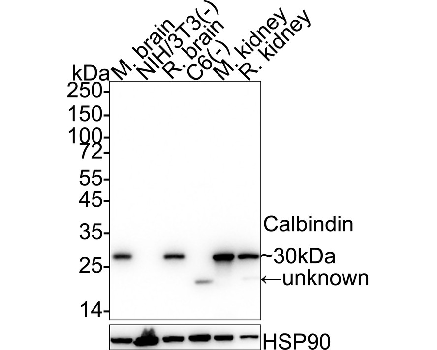
Western blot analysis of Calbindin on different lysates with Rabbit anti-Calbindin antibody at 1/5,000 dilution. Lane 1: Mouse brain tissue lysate, Lane 2: NIH/3T3 cell lysate (negative), Lane 3: Rat brain tissue lysate, Lane 4: C6 cell lysate (negative), Lane 5: Mouse kidney tissue lysate, Lane 6: Rat kidney tissue lysate, Lysates/proteins at 20 µg/Lane. Exposure time: 6 seconds; 4-20% SDS-PAGE gel. Proteins were transferred to a PVDF membrane and blocked with 5% NFDM/TBST for 1 hour at room temperature. The primary antibody at 1/5,000 dilution was used in 5% NFDM/TBST at 4℃ overnight. Goat Anti-Rabbit IgG - HRP Secondary Antibody at 1/50,000 dilution was used for 1 hour at room temperature.IHC
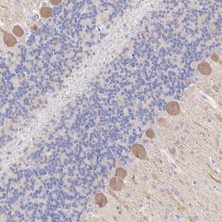
Immunohistochemical analysis of paraffin-embedded rat cerebellum tissue with Rabbit anti-Calbindin antibody at 1/10,000 dilution. The section was pre-treated using heat mediated antigen retrieval with Tris-EDTA buffer (pH 9.0) for 20 minutes. The tissues were blocked in 1% BSA for 20 minutes at room temperature, washed with ddH2O and PBS, and then probed with the primary antibody at 1/10,000 dilution for 1 hour at room temperature. The detection was performed using an HRP conjugated compact polymer system. DAB was used as the chromogen. Tissues were counterstained with hematoxylin and mounted with DPX.IHC
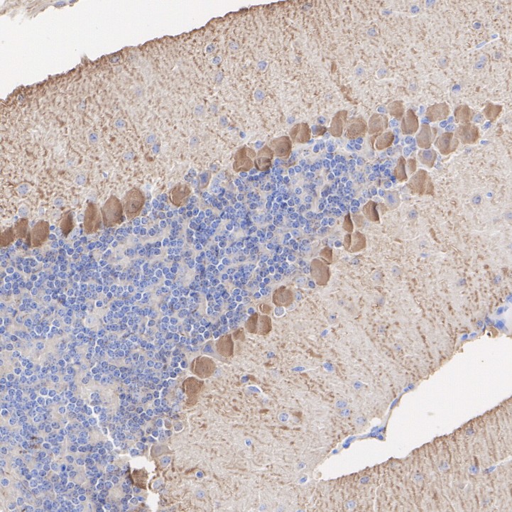
Immunohistochemical analysis of paraffin-embedded mouse cerebellum tissue with Rabbit anti-Calbindin antibody at 1/10,000 dilution. The section was pre-treated using heat mediated antigen retrieval with Tris-EDTA buffer (pH 9.0) for 20 minutes. The tissues were blocked in 1% BSA for 20 minutes at room temperature, washed with ddH2O and PBS, and then probed with the primary antibody at 1/10,000 dilution for 1 hour at room temperature. The detection was performed using an HRP conjugated compact polymer system. DAB was used as the chromogen. Tissues were counterstained with hematoxylin and mounted with DPX.IF-P
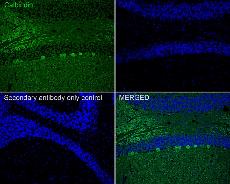
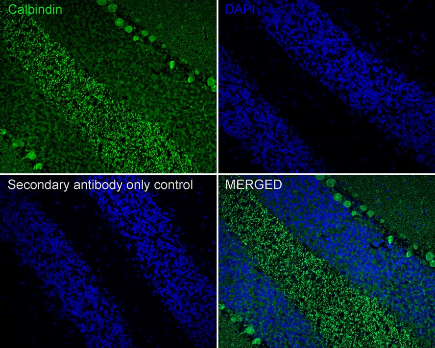
Species: Mouse, Site: Cerebellum, Sample: Paraffin-embedded section, Antibody concentration: 1/500.IF-P
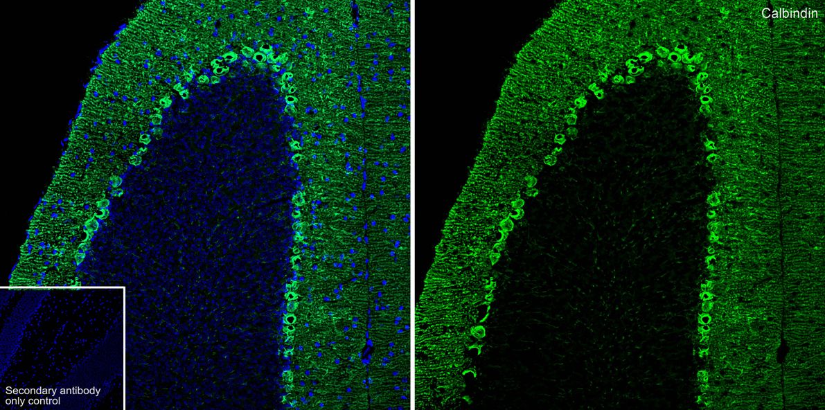
Species: Rat, Site: Cerebellum, Sample: Paraffin-embedded section, Antibody concentration: 1/500.IF-F
Species: Mouse, Site: Cerebellum, Sample: Frozen section, Antibody concentration: 1/2,000, Antigen retrieval: Not required.| Product Name | Calbindin Recombinant Rabbit Monoclonal Antibody |
|---|---|
| Antibody Type | Primary Antibodies |
| Product description | The family of EF-hand type Ca;2+;-binding proteins includes Calbindin D28K, Calbindin D9K, S-100 α and β, Calgranulin A (also designated MRP8), Calgranulin B (also designated MRP14), Calgranulin C and the Parvalbumin family members, including Parvalbumin α and Parvalbumin β (also designated oncomodulin). Calbindin D28K, also known as calbindin, CALB1, D-28K or vitamin D-dependent calcium-binding protein, is a 261 amino acid protein with six EF-hand domains, four of which are active calcium-binding domains. Expressed in brain, ovary, uterus, testis, pancreas, liver, kidney and intestine, Calbindin D28K acts as a calcium-buffering agent and alters the activity of the plasma membrane ATPase. In neuronal cells, Calbindin D28K modulates calcium channel activity, calcium transients and intrinsic neuronal firing activity. Also, Calbindin D28K has been implicated to play a role in apoptosis and microtubule function. |
| Immunogen | Synthetic peptide within Human Calbindin aa 51-100 / 261. |
| Clonality | Monoclonal |
|---|---|
| Isotype | IgG |
| Host Species | Recombinant rabbit |
| Tested Applications | IF-FIF-PIHCWB |
| WB:1:1000-1:5000 IHC:1:5000-1:10000 IF-P:1:500 IF-F:1:2000 |
|
| Species Reactivity | MouseRat |
| Concentration | 1mg/ml |
| Purification | Protein A |
| Alternative Names | avian-type antibody CAB27 antibody CALB 1 antibody CALB antibody CALB1 antibody CALB1_HUMAN antibody Calbindin 1 28kDa antibody Calbindin antibody Calbindin D28 antibody D 28K antibody D-28K antibody D28K antibody OTTHUMP00000166027 antibody OTTHUMP00000225441 antibody RTVL H protein antibody Vitamin D dependent calcium binding protein antibody Vitamin D dependent calcium binding protein avian type antibody Vitamin D-dependent calcium-binding protein antibody |
|---|---|
| Molecular Weight(MW) | 30kDa |
| Cellular Localization | Cytosol, Nucleus. |
WB
Western blot analysis of Calbindin on different lysates with Rabbit anti-Calbindin antibody at 1/5,000 dilution. Lane 1: Mouse brain tissue lysate, Lane 2: NIH/3T3 cell lysate (negative), Lane 3: Rat brain tissue lysate, Lane 4: C6 cell lysate (negative), Lane 5: Mouse kidney tissue lysate, Lane 6: Rat kidney tissue lysate, Lysates/proteins at 20 µg/Lane. Exposure time: 6 seconds; 4-20% SDS-PAGE gel. Proteins were transferred to a PVDF membrane and blocked with 5% NFDM/TBST for 1 hour at room temperature. The primary antibody at 1/5,000 dilution was used in 5% NFDM/TBST at 4℃ overnight. Goat Anti-Rabbit IgG - HRP Secondary Antibody at 1/50,000 dilution was used for 1 hour at room temperature.
IHC
Immunohistochemical analysis of paraffin-embedded rat cerebellum tissue with Rabbit anti-Calbindin antibody at 1/10,000 dilution. The section was pre-treated using heat mediated antigen retrieval with Tris-EDTA buffer (pH 9.0) for 20 minutes. The tissues were blocked in 1% BSA for 20 minutes at room temperature, washed with ddH2O and PBS, and then probed with the primary antibody at 1/10,000 dilution for 1 hour at room temperature. The detection was performed using an HRP conjugated compact polymer system. DAB was used as the chromogen. Tissues were counterstained with hematoxylin and mounted with DPX.
IHC
Immunohistochemical analysis of paraffin-embedded mouse cerebellum tissue with Rabbit anti-Calbindin antibody at 1/10,000 dilution. The section was pre-treated using heat mediated antigen retrieval with Tris-EDTA buffer (pH 9.0) for 20 minutes. The tissues were blocked in 1% BSA for 20 minutes at room temperature, washed with ddH2O and PBS, and then probed with the primary antibody at 1/10,000 dilution for 1 hour at room temperature. The detection was performed using an HRP conjugated compact polymer system. DAB was used as the chromogen. Tissues were counterstained with hematoxylin and mounted with DPX.
IF-P
Species: Mouse, Site: Cerebellum, Sample: Paraffin-embedded section, Antibody concentration: 1/500.
IF-P
Species: Rat, Site: Cerebellum, Sample: Paraffin-embedded section, Antibody concentration: 1/500.
IF-F
Species: Mouse, Site: Cerebellum, Sample: Frozen section, Antibody concentration: 1/2,000, Antigen retrieval: Not required.| Application Notes | WB:1:1000-1:5000 IHC:1:5000-1:10000 IF-P:1:500 IF-F:1:2000 |
|---|
| Form | Liquid |
|---|---|
| Storage Instructions | Store at +4℃ after thawing. Aliquot store at -20℃ or -80℃. Avoid repeated freeze / thaw cycles. |
| Storage Buffer | 1*TBS (pH7.4), 1%BSA, 40%Glycerol. Preservative: 0.05% Sodium Azide. |


