Application
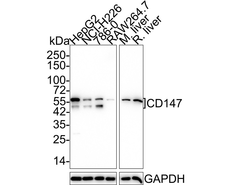
Fig1:Western blot analysis of CD147 on different lysates with Rabbit anti-CD147 antibody at 1/5,000 dilution. Lane 1: HepG2 cell lysate (15 µg/Lane), Lane 2: NCI-H226 cell lysate (15 µg/Lane), Lane 3: 786-0 cell lysate (15 µg/Lane), Lane 4: RAW264.7 cell lysate (15 µg/Lane), Lane 5: Mouse liver tissue lysate (20 µg/Lane), Lane 6: Rat liver tissue lysate (20 µg/Lane), Exposure time: 3 minutes; 4-20% SDS-PAGE gel. Proteins were transferred to a PVDF membrane and blocked with 5% NFDM/TBST for 1 hour at room temperature. The primary antibody at 1/5,000 dilution was used in 5% NFDM/TBST at 4℃ overnight. Goat Anti-Rabbit IgG - HRP Secondary Antibody at 1/50,000 dilution was used for 1 hour at room temperature.Application
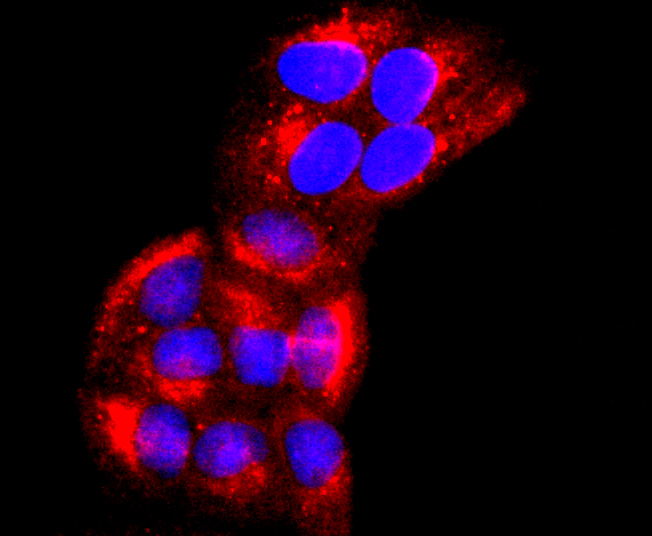
Fig2: ICC staining CD147 in Hela cells (red). The nuclear counter stain is DAPI (blue). Cells were fixed in paraformaldehyde, permeabilised with 0.25% Triton X100/PBS.Application
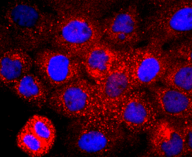
Fig3: ICC staining CD147 in A431 cells (red). The nuclear counter stain is DAPI (blue). Cells were fixed in paraformaldehyde, permeabilised with 0.25% Triton X100/PBS.Application
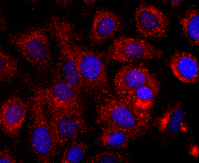
Fig4: ICC staining CD147 in SKOV-3 cells (red). The nuclear counter stain is DAPI (blue). Cells were fixed in paraformaldehyde, permeabilised with 0.25% Triton X100/PBS.Application
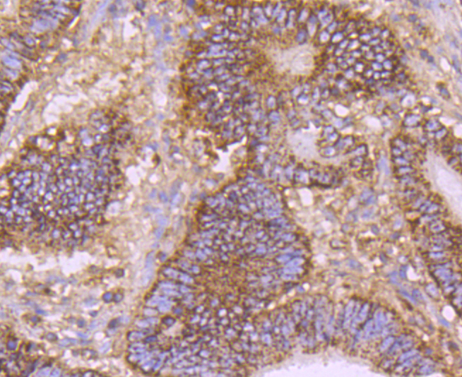
Fig5: Immunohistochemical analysis of paraffin-embedded human colon cancer tissue using anti-CD147 antibody. Counter stained with hematoxylin.Application
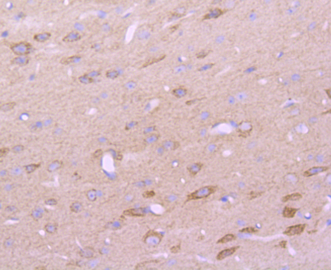
Fig6: Immunohistochemical analysis of paraffin-embedded mouse brain tissue using anti-CD147 antibody. Counter stained with hematoxylin.Application
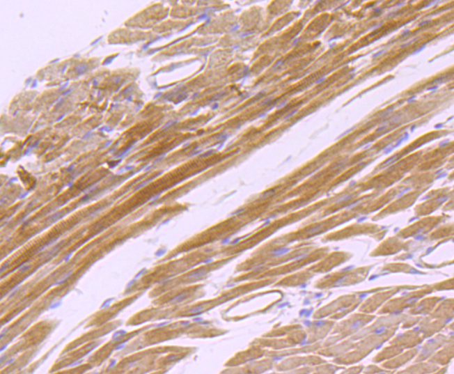
Fig7: Immunohistochemical analysis of paraffin-embedded mouse heart tissue using anti-CD147 antibody. Counter stained with hematoxylin.Application
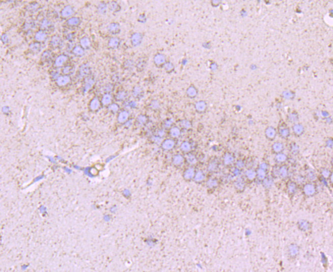
Fig8: Immunohistochemical analysis of paraffin-embedded rat brain tissue using anti-CD147 antibody. Counter stained with hematoxylin.Application
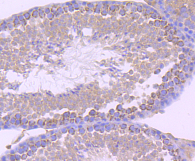
Fig9: Immunohistochemical analysis of paraffin-embedded mouse testis tissue using anti-CD147 antibody. Counter stained with hematoxylin.Application
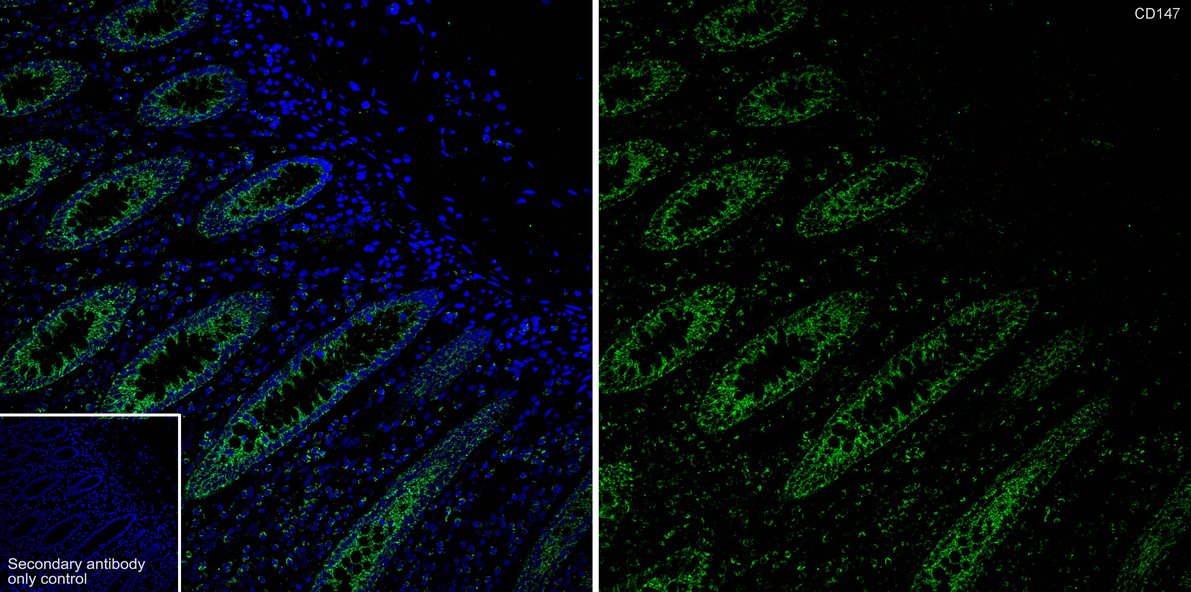
Fig10: Immunofluorescence analysis of paraffin-embedded human colon tissue labeling CD147 with Rabbit anti-CD147 antibody at 1/200 dilution. The section was pre-treated using heat mediated antigen retrieval with Tris-EDTA buffer (pH 9.0) for 20 minutes. The tissues were blocked in 10% negative goat serum for 1 hour at room temperature, washed with PBS, and then probed with the primary antibody (green) at 1/200 dilution overnight at 4 ℃, washed with PBS. Goat Anti-Rabbit IgG H&L (488) was used as the secondary antibody at 1/1,000 dilution. Nuclei were counterstained with DAPI (blue).| Product Name | CD147 Recombinant Rabbit Monoclonal Antibody |
|---|---|
| Antibody Type | Primary Antibodies |
| Product description | Extracellular matrix metalloproteinase inducer (EMMPRIN), also designated basigin or CD147, is involved in the regulation of matrix remodeling at the epidermal-dermal interface. EMMPRIN stimulates the production of interstitial collagenase, gelatinase A, stromelysin-1 and various metalloproteinases (MMPs) by fibroblasts. These enzymes, which are typically increased during tissue degradation and wound healing, are important factors in cancer invasion and metastasis. |
| Immunogen | Synthetic peptide within Human CD147 aa 171-204 / 385. |
| Clonality | Monoclonal |
|---|---|
| Isotype | IgG |
| Host Species | Recombinant rabbit |
| Tested Applications | ICC/IFIF-PIHCWB |
| WB:1:5000 ICC/IF:1:50-1:200 IHC:1:50-1:200 IF-P:1:50-1:200 |
|
| Species Reactivity | HumanMouseRat |
| Concentration | 1mg/ml |
| Purification | Protein A |
| Alternative Names | 5A11 antigen antibody 5F7 antibody BASI_HUMAN antibody Basigin (Ok blood group) antibody Basigin antibody Blood brain barrier HT7 antigen antibody Bsg antibody CD 147 antibody CD147 antibody CD147 antigen antibody Collagenase stimulatory factor antibody EMMPRIN antibody Extracellular matrix metalloproteinase inducer antibody Leukocyte activation antigen M6 antibody M 6 antibody M6 antibody M6 leukocyte activation antigen antibody Neurothelin antibody OK antibody OK blood group antibody OK blood group antigen antibody TCSF antibody Tumor cell derived collagenase stimulatory factor antibody Tumor cell-derived collagenase stimulatory factor antibody |
|---|---|
| Molecular Weight(MW) | 42kDa(Observed band size: 45-60kDa) |
| Cellular Localization | Cell membrane, Melanosome. |
Application
Fig1:Western blot analysis of CD147 on different lysates with Rabbit anti-CD147 antibody at 1/5,000 dilution. Lane 1: HepG2 cell lysate (15 µg/Lane), Lane 2: NCI-H226 cell lysate (15 µg/Lane), Lane 3: 786-0 cell lysate (15 µg/Lane), Lane 4: RAW264.7 cell lysate (15 µg/Lane), Lane 5: Mouse liver tissue lysate (20 µg/Lane), Lane 6: Rat liver tissue lysate (20 µg/Lane), Exposure time: 3 minutes; 4-20% SDS-PAGE gel. Proteins were transferred to a PVDF membrane and blocked with 5% NFDM/TBST for 1 hour at room temperature. The primary antibody at 1/5,000 dilution was used in 5% NFDM/TBST at 4℃ overnight. Goat Anti-Rabbit IgG - HRP Secondary Antibody at 1/50,000 dilution was used for 1 hour at room temperature.
Application
Fig2: ICC staining CD147 in Hela cells (red). The nuclear counter stain is DAPI (blue). Cells were fixed in paraformaldehyde, permeabilised with 0.25% Triton X100/PBS.
Application
Fig3: ICC staining CD147 in A431 cells (red). The nuclear counter stain is DAPI (blue). Cells were fixed in paraformaldehyde, permeabilised with 0.25% Triton X100/PBS.
Application
Fig4: ICC staining CD147 in SKOV-3 cells (red). The nuclear counter stain is DAPI (blue). Cells were fixed in paraformaldehyde, permeabilised with 0.25% Triton X100/PBS.
Application
Fig5: Immunohistochemical analysis of paraffin-embedded human colon cancer tissue using anti-CD147 antibody. Counter stained with hematoxylin.
Application
Fig6: Immunohistochemical analysis of paraffin-embedded mouse brain tissue using anti-CD147 antibody. Counter stained with hematoxylin.
Application
Fig7: Immunohistochemical analysis of paraffin-embedded mouse heart tissue using anti-CD147 antibody. Counter stained with hematoxylin.
Application
Fig8: Immunohistochemical analysis of paraffin-embedded rat brain tissue using anti-CD147 antibody. Counter stained with hematoxylin.
Application
Fig9: Immunohistochemical analysis of paraffin-embedded mouse testis tissue using anti-CD147 antibody. Counter stained with hematoxylin.
Application
Fig10: Immunofluorescence analysis of paraffin-embedded human colon tissue labeling CD147 with Rabbit anti-CD147 antibody at 1/200 dilution. The section was pre-treated using heat mediated antigen retrieval with Tris-EDTA buffer (pH 9.0) for 20 minutes. The tissues were blocked in 10% negative goat serum for 1 hour at room temperature, washed with PBS, and then probed with the primary antibody (green) at 1/200 dilution overnight at 4 ℃, washed with PBS. Goat Anti-Rabbit IgG H&L (488) was used as the secondary antibody at 1/1,000 dilution. Nuclei were counterstained with DAPI (blue).| Application Notes | WB:1:5000 ICC/IF:1:50-1:200 IHC:1:50-1:200 IF-P:1:50-1:200 |
|---|
| Form | Liquid |
|---|---|
| Storage Instructions | Store at +4℃ after thawing. Aliquot store at -20℃ or -80℃. Avoid repeated freeze / thaw cycles. |
| Storage Buffer | 1*TBS (pH7.4), 1%BSA, 40%Glycerol. Preservative: 0.05% Sodium Azide. |


