ICC/IF
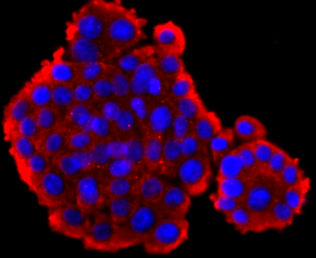
ICC staining GLP1 in PC-12 cells (red). The nuclear counter stain is DAPI (blue). Cells were fixed in paraformaldehyde, permeabilised with 0.25% Triton X100/PBS.ICC/IF
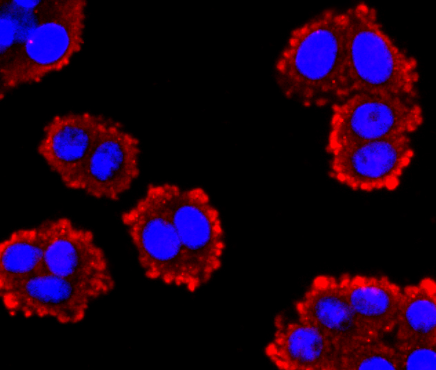
ICC staining GLP1 in SW480 cells (red). The nuclear counter stain is DAPI (blue). Cells were fixed in paraformaldehyde, permeabilised with 0.25% Triton X100/PBS.IHC
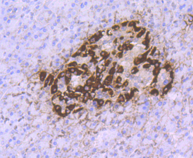
Immunohistochemical analysis of paraffin-embedded human pancreas tissue using anti-GLP1 antibody. Counter stained with hematoxylin.IF-P
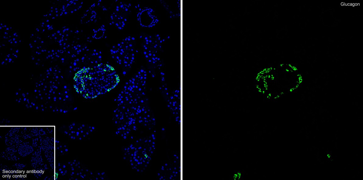
Immunofluorescence analysis of paraffin-embedded mouse pancreas tissue labeling Glucagon with Rabbit anti-Glucagon antibody at 1/100 dilution. The section was pre-treated using heat mediated antigen retrieval with Tris-EDTA buffer (pH 9.0) for 20 minutes. The tissues were blocked in 10% negative goat serum for 1 hour at room temperature, washed with PBS, and then probed with the primary antibody ( green) at 1/100 dilution overnight at 4 ℃, washed with PBS. Goat Anti-Rabbit IgG H&L (488) was used as the secondary antibody at 1/1,000 dilution. Nuclei were counterstained with DAPI (blue).| Product Name | Glucagon Recombinant Rabbit Monoclonal Antibody |
|---|---|
| Antibody Type | Primary Antibodies |
| Product description | Glucagon is a pancreatic hormone that functions as an antagonist to insulin, stimulating the conversion of glycogen to glucose and increasing blood sugar levels. Glucagon-like peptide-1 (GLP-1), Glucagon-like peptide-2 (GLP-2), VIP (vasoactive intestinal peptide) and PACAP (pituitary adenylate cyclase activating polypeptide) are members of the Glucagon family of hormones. GLP-1 functions as a transmitter in the central nervous system, inhibiting feeding and drinking behavior, whereas GLP-2 is a stimulator of intestinal epithelial growth. VIP causes vasodilation resulting in the lowering of blood pressure. PACAP is abundant in the hypothalamus and has been shown to increase the synthesis of several hormones, including growth hormone. |
| Immunogen | Recombinant protein within human GLP1 aa 65-150. |
| Clonality | Monoclonal |
|---|---|
| Isotype | IgG |
| Host Species | Recombinant rabbit |
| Tested Applications | ICC/IFIF-PIHCWB |
| WB:1 500-1:1000ICC/IF:1:50-1:200 IHC:1:50-1:200 IF-P:1:100 |
|
| Species Reactivity | HumanMouseRat |
| Concentration | 1mg/ml |
| Purification | Protein A |
| Alternative Names | GCG antibody
glicentin-related polypeptide antibody
GLP-1 antibody
GLP-1(7-36) antibody
GLP-1(7-37) antibody
GLP-2 antibody
GLP1 antibody
GLP1 included antibody GLP2 antibody GLP2 included antibody GLUC_HUMAN antibody Glucagon antibody Glucagon like peptide 1 antibody glucagon-like peptide 1 antibody Glucagon-like peptide 1 included antibody Glucagon-like peptide 2 antibody Glucagon-like peptide 2 included antibody GRPP antibody OXM antibody OXY antibody preproglucagon antibody |
|---|---|
| Molecular Weight(MW) | 21/8/4 kDa |
| Cellular Localization | Secreted. |
| SwissProt ID | P01275 |
|---|
ICC/IF
ICC staining GLP1 in PC-12 cells (red). The nuclear counter stain is DAPI (blue). Cells were fixed in paraformaldehyde, permeabilised with 0.25% Triton X100/PBS.
ICC/IF
ICC staining GLP1 in SW480 cells (red). The nuclear counter stain is DAPI (blue). Cells were fixed in paraformaldehyde, permeabilised with 0.25% Triton X100/PBS.
IHC
Immunohistochemical analysis of paraffin-embedded human pancreas tissue using anti-GLP1 antibody. Counter stained with hematoxylin.
IF-P
Immunofluorescence analysis of paraffin-embedded mouse pancreas tissue labeling Glucagon with Rabbit anti-Glucagon antibody at 1/100 dilution. The section was pre-treated using heat mediated antigen retrieval with Tris-EDTA buffer (pH 9.0) for 20 minutes. The tissues were blocked in 10% negative goat serum for 1 hour at room temperature, washed with PBS, and then probed with the primary antibody ( green) at 1/100 dilution overnight at 4 ℃, washed with PBS. Goat Anti-Rabbit IgG H&L (488) was used as the secondary antibody at 1/1,000 dilution. Nuclei were counterstained with DAPI (blue).| Application Notes | WB:1 500-1:1000ICC/IF:1:50-1:200 IHC:1:50-1:200 IF-P:1:100 |
|---|
| Form | Liquid |
|---|---|
| Storage Instructions | Store at +4℃ after thawing. Aliquot store at -20℃ or -80℃. Avoid repeated freeze / thaw cycles. |
| Storage Buffer | 1*TBS (pH7.4), 1%BSA, 40%Glycerol. Preservative: 0.05% Sodium Azide. |


