Application
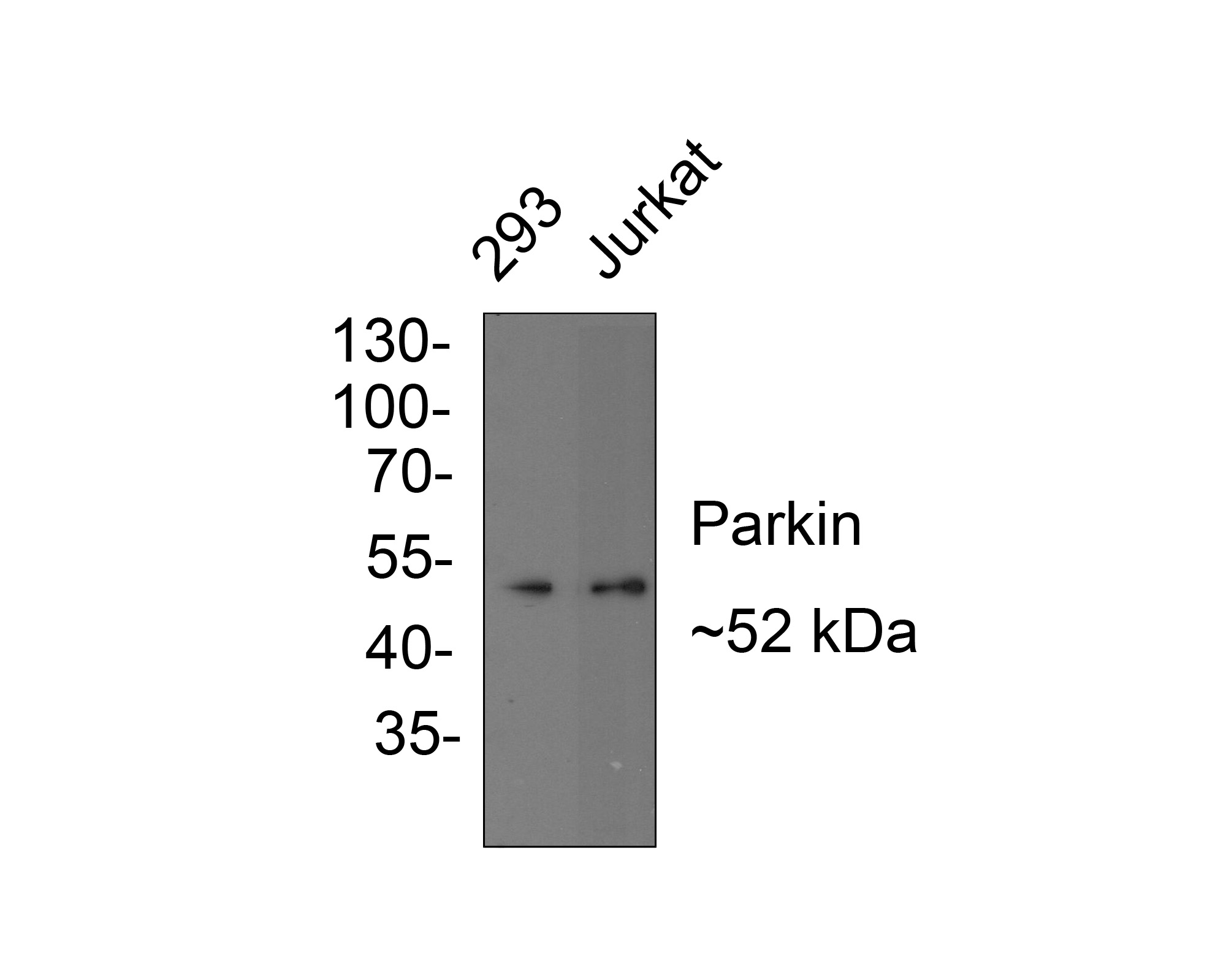
Fig1: Western blot analysis of Parkin on different lysates with Rabbit anti-Parkin antibody at 1/1,000 dilution. Lane 1: 293 cell lysate, Lane 2: Jurkat cell lysate, Lysates/proteins at 10 µg/Lane. Exposure time: 1 minutes; 10% SDS-PAGE gel. Proteins were transferred to a PVDF membrane and blocked with 5% NFDM/TBST for 1 hour at room temperature. The primary antibody at 1/1,000 dilution was used in 5% NFDM/TBST at room temperature for 2 hours. Goat Anti-Rabbit IgG - HRP Secondary Antibody at 1:300,000 dilution was used for 1 hour at room temperature.Application
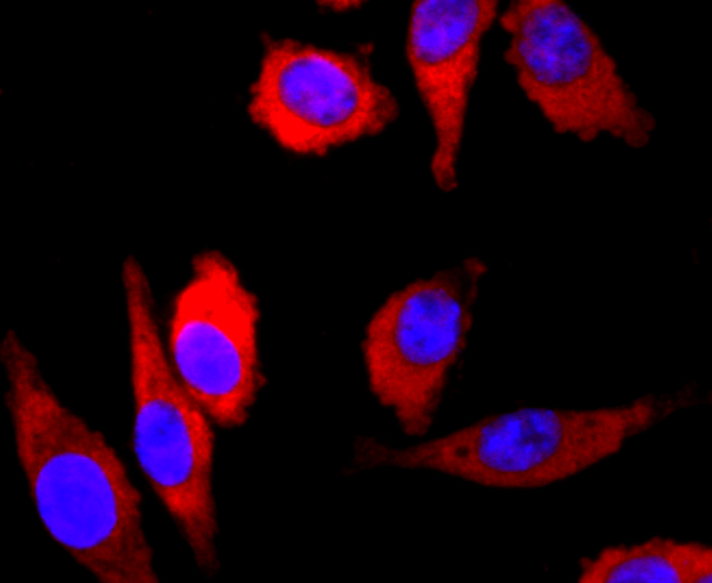
Fig2: ICC staining Parkin in SH-SY-5Y cells (red). The nuclear counter stain is DAPI (blue). Cells were fixed in paraformaldehyde, permeabilised with 0.25% Triton X100/PBS.Application
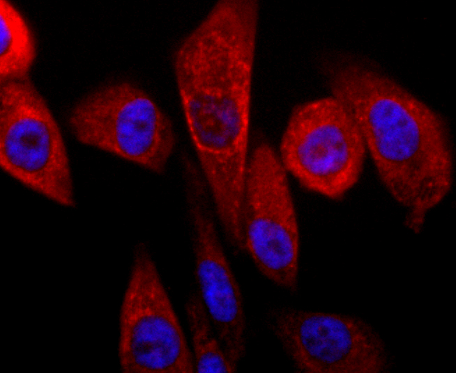
Fig3: ICC staining Parkin in PC-3M cells (red). The nuclear counter stain is DAPI (blue). Cells were fixed in paraformaldehyde, permeabilised with 0.25% Triton X100/PBS.Application
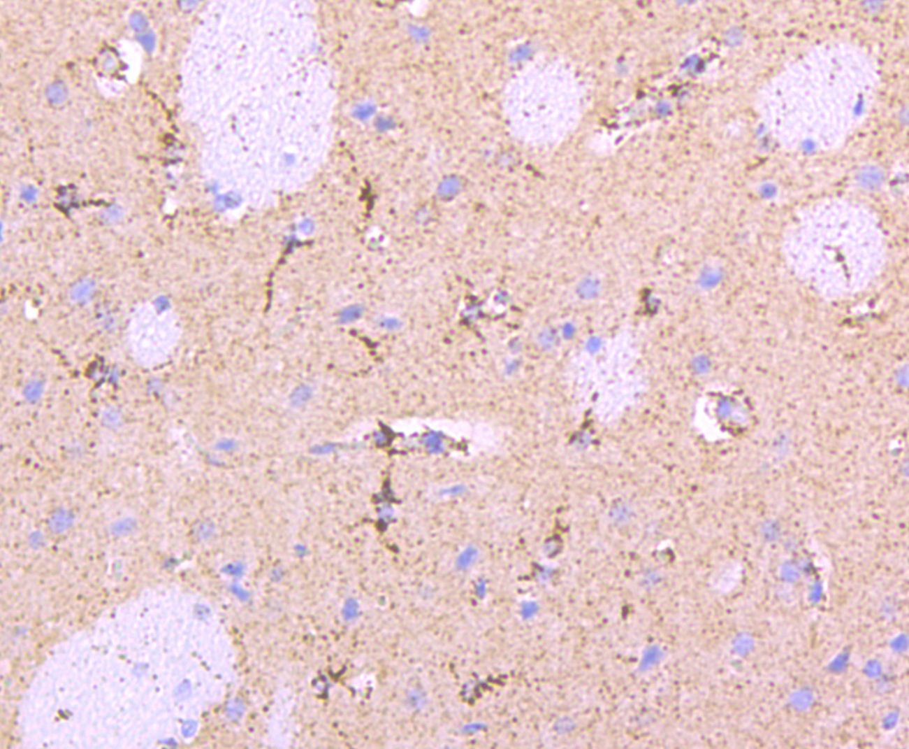
Fig4: Immunohistochemical analysis of paraffin-embedded rat brain tissue using anti-Parkin antibody. Counter stained with hematoxylin.Application
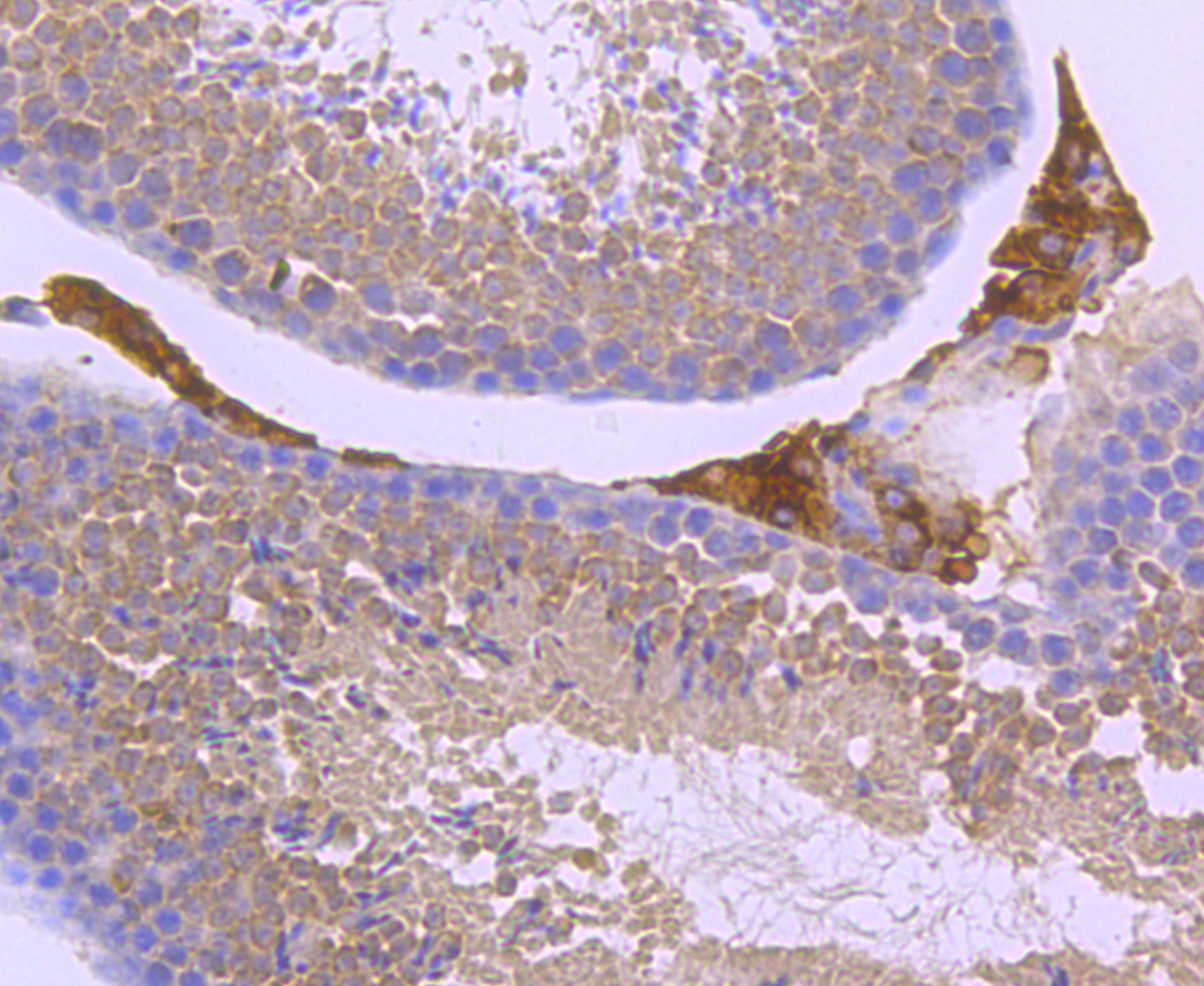
Fig5: Immunohistochemical analysis of paraffin-embedded mouse testis tissue using anti-Parkin antibody. Counter stained with hematoxylin.Application
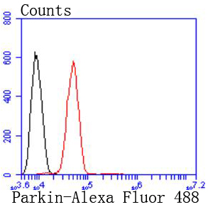
Fig6: Flow cytometric analysis of SH-SY-5Y cells with Parkin antibody at 1/50 dilution (red) compared with an unlabelled control (cells without incubation with primary antibody; black). Alexa Fluor 488-conjugated goat anti rabbit IgG was used as the secondary antibody| Product Name | Parkin Recombinant Rabbit Monoclonal Antibody |
|---|---|
| Antibody Type | Primary Antibodies |
| Product description | Parkin is a zinc-finger protein that is related to ubiquitin at the amino terminus. The wild type Parkin gene, which maps to human chromosome 6q25.2-27, encodes a 465 amino acid full-length protein that is expressed as multiple isoforms. Mutations in the Parkin gene are responsible for autosomal recessive juvenile Parkinson's disease and commonly involve deletions of exons 3-5. In humans, Parkin is expressed in a subset of cells of the basal ganglia, midbrain, cerebellum and cerebral cortex, and is subject to alternative splicing in different tissues. Parkin expression is also high in the brainstem of mice, with the majority of immunopositive cells being neurons. The Parkin gene has been identified in a diverse group of organisms including mammals, birds, frog and fruit flies, suggesting that analogous functional roles of the Parkin protein may have been highly conserved during the course of evolution. |
| Immunogen | Synthetic peptide within N-terminal human Parkin. |
| Clonality | monoclonal |
|---|---|
| Isotype | IgG |
| Host Species | Recombinant rabbit |
| Tested Applications | FCICC/IFIHCIPWB |
| WB:1:500-1:1,000 ICC:1:50-1:200 IHC:1:50-1:200 FC:1:50-1:100 IP:1:50 |
|
| Species Reactivity | HumanMouseRat |
| Concentration | 1mg/ml |
| Purification | Protein A |
| Alternative Names | AR JP antibody
E3 ubiquitin ligase antibody
E3 ubiquitin protein ligase parkin antibody
E3 ubiquitin-protein ligase parkin antibody
FRA6E antibody
LPRS 2 antibody
LPRS2 antibody
PARK 2 antibody
Park2 antibody
Parkin 2 antibody
Parkinson disease (autosomal recessive juvenile) 2 antibody
Parkinson disease (autosomal recessive juvenile) 2 parkin antibody Parkinson disease protein 2 antibody Parkinson juvenile disease protein 2 antibody Parkinson protein 2 E3 ubiquitin protein ligase antibody Parkinson protein 2 E3 ubiquitin protein ligase (parkin) antibody PDJ antibody PRKN 2 antibody PRKN antibody PRKN2 antibody PRKN2_HUMAN antibody Ubiquitin E3 ligase PRKN antibody |
|---|---|
| Molecular Weight(MW) | 52kDa |
| Cellular Localization | Cytoplasm, Nucleus, Endoplasmic reticulum, Mitochondrion. |
Application
Fig1: Western blot analysis of Parkin on different lysates with Rabbit anti-Parkin antibody at 1/1,000 dilution. Lane 1: 293 cell lysate, Lane 2: Jurkat cell lysate, Lysates/proteins at 10 µg/Lane. Exposure time: 1 minutes; 10% SDS-PAGE gel. Proteins were transferred to a PVDF membrane and blocked with 5% NFDM/TBST for 1 hour at room temperature. The primary antibody at 1/1,000 dilution was used in 5% NFDM/TBST at room temperature for 2 hours. Goat Anti-Rabbit IgG - HRP Secondary Antibody at 1:300,000 dilution was used for 1 hour at room temperature.
Application
Fig2: ICC staining Parkin in SH-SY-5Y cells (red). The nuclear counter stain is DAPI (blue). Cells were fixed in paraformaldehyde, permeabilised with 0.25% Triton X100/PBS.
Application
Fig3: ICC staining Parkin in PC-3M cells (red). The nuclear counter stain is DAPI (blue). Cells were fixed in paraformaldehyde, permeabilised with 0.25% Triton X100/PBS.
Application
Fig4: Immunohistochemical analysis of paraffin-embedded rat brain tissue using anti-Parkin antibody. Counter stained with hematoxylin.
Application
Fig5: Immunohistochemical analysis of paraffin-embedded mouse testis tissue using anti-Parkin antibody. Counter stained with hematoxylin.
Application
Fig6: Flow cytometric analysis of SH-SY-5Y cells with Parkin antibody at 1/50 dilution (red) compared with an unlabelled control (cells without incubation with primary antibody; black). Alexa Fluor 488-conjugated goat anti rabbit IgG was used as the secondary antibody| Positive Control | N2A, SH-SY-5Y, PC-3M, rat brain tissue, mouse testis tissue. |
|---|---|
| Application Notes | WB:1:500-1:1,000 ICC:1:50-1:200 IHC:1:50-1:200 FC:1:50-1:100 IP:1:50 |
| Form | Liquid |
|---|---|
| Storage Instructions | Store at +4℃ after thawing. Aliquot store at -20℃ or -80℃. Avoid repeated freeze / thaw cycles. |
| Storage Buffer | 1*TBS (pH7.4), 1%BSA, 40%Glycerol. Preservative: 0.05% Sodium Azide. |


