Application
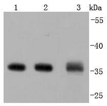
Fig1: Western blot analysis of Annexin V on different lysates using anti-Annexin V antibody at 1/1,000 dilution. Positive control: Lane 1: Hela Lane 2: HepG2 Lane 3: JARApplication
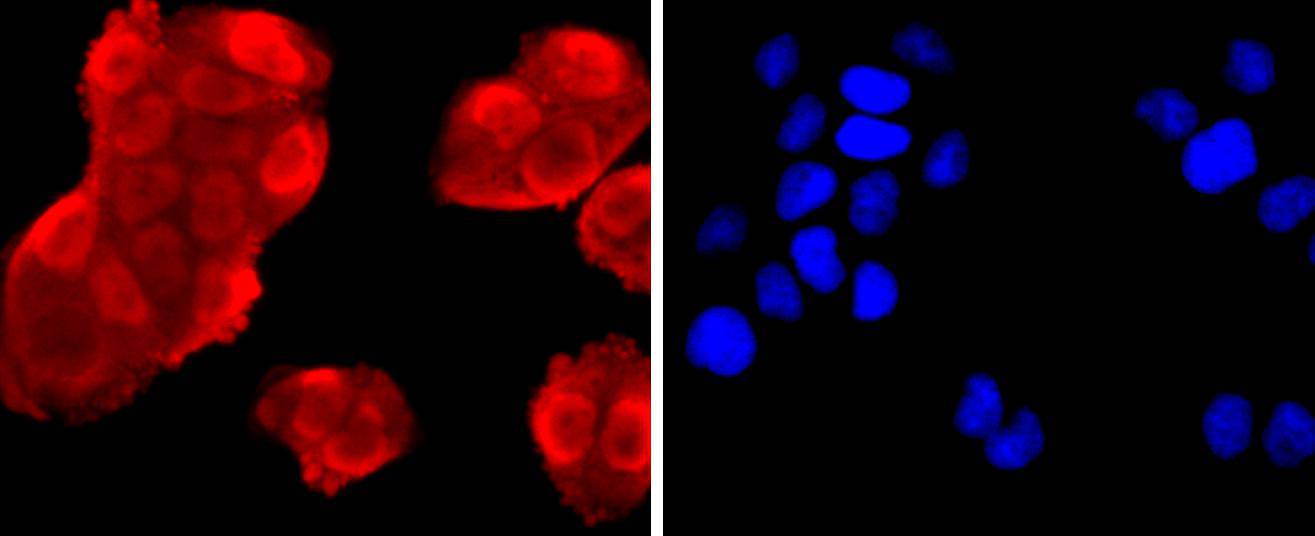
Fig2: ICC staining Annexin V in Hela cells (red). The nuclear counter stain is DAPI (blue). Cells were fixed in paraformaldehyde, permeabilised with 0.25% Triton X100/PBS.Application
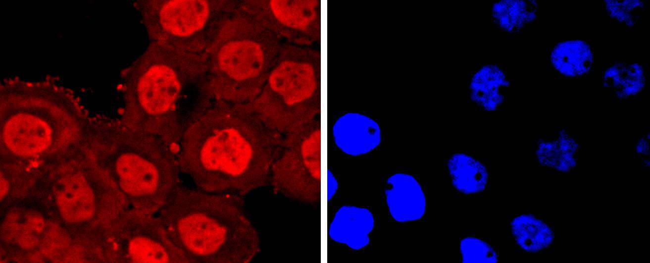
Fig3: ICC staining Annexin V in A431 cells (red). The nuclear counter stain is DAPI (blue). Cells were fixed in paraformaldehyde, permeabilised with 0.25% Triton X100/PBS.Application
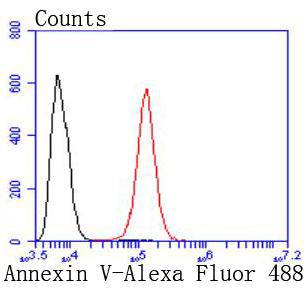
Fig4: Flow cytometric analysis of Hela cells with Annexin V antibody at 1/50 dilution (red) compared with an unlabelled control (cells without incubation with primary antibody; black). Alexa Fluor 488-conjugated goat anti rabbit IgG was used as the secondary antibody| Product Name | Annexin V [JF50-11] |
|---|---|
| Antibody Type | Primary Antibodies |
| Antigen Alias | Anchorin CII antibody Annexin 5 antibody Annexin A5 antibody Annexin V antibody Annexin-5 antibody ANX A5 antibody ANX5 antibody ANXA5 antibody ANXA5_HUMAN antibody Calphobindin I antibody CBP-I antibody Endonexin II antibody ENX2 antibody Lipocortin V antibody PAP I antibody PAP-I antibody Placental anticoagulant protein 4 antibody Placental anticoagulant protein I antibody PLACENTAL PROTEIN 4 antibody Pp4 antibody Thromboplastin inhibitor antibody VAC-alpha antibody Vascular anticoagulant-alpha antibody |
| Product description | The annexin family of calcium-binding proteins is composed of at least ten mammalian genes and is characterized by a conserved core domain, which binds phospholipids in a Ca;2+;-dependent manner, and a unique amino-terminal region, which may confer binding specificity. Annexin family members have been implicated as regulators of such diverse processes as ion flux, endocytosis and exocytosis, and cellular adhesion. For example, the crystal structure of Annexin III has suggested a hydrophilic amino-terminus with possible Ca;2+; channel activity. Similarly, Annexin V has ion channel properties. Annexin IV, also referred to as endonexin, functions to regulate Cl-flux by mediating calmodulin kinase II (CaMKII) activity and Annexin V has been shown to regulate PKC activity. Annexin V is ubiquitously expressed at high levels in tissues and cells grown in tissue culture, while Annexin VIII exhibits a more limited distribution. Where co-expressed in the same tissues, Annexin VIII is often expressed at a 100-fold lower level than Annexin V. However, Annexin VIII is preferentially expressed in acute promyelocytic leukemia (APL) cells, which may relate to its role in hematopoietic cell differentiation. |
| Immunogen | recombinant protein |
| Clonality | Monoclonal |
|---|---|
| Isotype | IgG |
| Host Species | Recombinant rabbit |
| Tested Applications | WBICC/IFFC |
| WB:1:1,000 FC:1:50-1:100 ICC:1:100-1:500 |
|
| Species Reactivity | HumanMouseRat |
| Concentration | 1mg/ml |
| Alternative Names | Anchorin CII antibody Annexin 5 antibody Annexin A5 antibody Annexin V antibody Annexin-5 antibody ANX A5 antibody ANX5 antibody ANXA5 antibody ANXA5_HUMAN antibody Calphobindin I antibody CBP-I antibody Endonexin II antibody ENX2 antibody Lipocortin V antibody PAP I antibody PAP-I antibody Placental anticoagulant protein 4 antibody Placental anticoagulant protein I antibody PLACENTAL PROTEIN 4 antibody Pp4 antibody Thromboplastin inhibitor antibody VAC-alpha antibody Vascular anticoagulant-alpha antibody |
|---|---|
| Molecular Weight(MW) | 36 kDa |
| Cellular Localization | Cytoplasm, endothelial microparticle, membrane. |
Application
Fig1: Western blot analysis of Annexin V on different lysates using anti-Annexin V antibody at 1/1,000 dilution. Positive control: Lane 1: Hela Lane 2: HepG2 Lane 3: JAR
Application
Fig2: ICC staining Annexin V in Hela cells (red). The nuclear counter stain is DAPI (blue). Cells were fixed in paraformaldehyde, permeabilised with 0.25% Triton X100/PBS.
Application
Fig3: ICC staining Annexin V in A431 cells (red). The nuclear counter stain is DAPI (blue). Cells were fixed in paraformaldehyde, permeabilised with 0.25% Triton X100/PBS.
Application
Fig4: Flow cytometric analysis of Hela cells with Annexin V antibody at 1/50 dilution (red) compared with an unlabelled control (cells without incubation with primary antibody; black). Alexa Fluor 488-conjugated goat anti rabbit IgG was used as the secondary antibody| Positive Control | JAR, A431, Hela, HepG2, human liver tissue, mouse liver tissue, mouse brain tissue, human lung tissue. |
|---|---|
| Application Notes | WB:1:1,000 FC:1:50-1:100 ICC:1:100-1:500 |
| Form | Liquid |
|---|---|
| Storage Instructions | Store at +4℃ after thawing. Aliquot store at -20℃ or -80℃. Avoid repeated freeze / thaw cycles. |
| Storage Buffer | 1*TBS (pH7.4), 1%BSA, 40%Glycerol. Preservative: 0.05% Sodium Azide. |


