Application
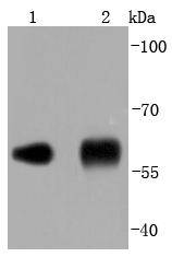
Fig1: Western blot analysis of PDI on different lysates using anti-PDI antibody at 1/1,000 dilution. Positive control: Lane 1: NIH/3T3 Lane 2: Human liverApplication
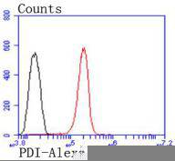
Fig2: Flow cytometric analysis of Hela cells with PDI antibody at 1/50 dilution (red) compared with an unlabelled control (cells without incubation with primary antibody; black). Alexa Fluor 488-conjugated goat anti rabbit IgG was used as the secondary antibody| Product Name | PDI [JF97-08] |
|---|---|
| Antibody Type | Primary Antibodies |
| Antigen Alias | Pancreas specific protein disulfide isomerase antibody Pancreas-specific protein disulfide isomerase antibody Pancreatic protein disulfide isomerase antibody PDA2 antibody PDI antibody PDIA2 antibody PDIA2_HUMAN antibody PDIp antibody PDIR antibody Protein disulfide isomerase A2 antibody Protein disulfide isomerase antibody Protein disulfide isomerase associated 2 antibody Protein disulfide isomerase family A member 2 antibody Protein disulfide isomerase pancreatic antibody Protein disulfide-isomerase A2 antibody Rho GDP dissociation inhibitor gamma antibody |
| Product description | Oxidoreductase-protein disulfide isomerase (PDI) is a homodimer consisting of subunits that catalyzes thiol-disulfide exchange, mediates folding of newly synthesized proteins and functions as a molecular chaperone. PDI localizes to the lumen of the endoplasmic reticulum (ER), where in conjunction with folding-helper proteins, such as immunoglobulin heavy chain binding protein (BiP), mediates tertiary and quaternary protein-processing. Cell surface PDI induces sulfhydryl-mediated conformational changes in integrin-mediated adhesion receptor-ligand interactions, thereby regulating integrin responses and cell adhesion. Additionally, PDI functions as a subunit of two more complex enzyme systems: the prolyl-4-hydroxylase and the triacylglycerol transfer proteins. |
| Immunogen | recombinant protein |
| Clonality | Monoclonal |
|---|---|
| Isotype | IgG |
| Host Species | Recombinant rabbit |
| Tested Applications | WBFC |
| WB:1:1,000-1:2,000 FC:1:50-1:100 |
|
| Species Reactivity | HumanMouseRat |
| Concentration | 1mg/ml |
| Alternative Names | Pancreas specific protein disulfide isomerase antibody Pancreas-specific protein disulfide isomerase antibody Pancreatic protein disulfide isomerase antibody PDA2 antibody PDI antibody PDIA2 antibody PDIA2_HUMAN antibody PDIp antibody PDIR antibody Protein disulfide isomerase A2 antibody Protein disulfide isomerase antibody Protein disulfide isomerase associated 2 antibody Protein disulfide isomerase family A member 2 antibody Protein disulfide isomerase pancreatic antibody Protein disulfide-isomerase A2 antibody Rho GDP dissociation inhibitor gamma antibody |
|---|---|
| Molecular Weight(MW) | 58 kDa |
| Cellular Localization | Endoplasmic reticulum, Endoplasmic reticulum lumen, Melanosome, Cell membrane. |
Application
Fig1: Western blot analysis of PDI on different lysates using anti-PDI antibody at 1/1,000 dilution. Positive control: Lane 1: NIH/3T3 Lane 2: Human liver
Application
Fig2: Flow cytometric analysis of Hela cells with PDI antibody at 1/50 dilution (red) compared with an unlabelled control (cells without incubation with primary antibody; black). Alexa Fluor 488-conjugated goat anti rabbit IgG was used as the secondary antibody| Positive Control | NIH/3T3, human liver tissue. |
|---|---|
| Application Notes | WB:1:1,000-1:2,000 FC:1:50-1:100 |
| Form | Liquid |
|---|---|
| Storage Instructions | Store at +4℃ after thawing. Aliquot store at -20℃ or -80℃. Avoid repeated freeze / thaw cycles. |
| Storage Buffer | 1*TBS (pH7.4), 1%BSA, 40%Glycerol. Preservative: 0.05% Sodium Azide. |


