Application
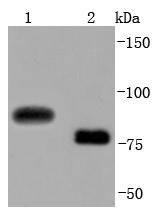
Fig1: Western blot analysis of CD19 on different lysates using anti-CD19 antibody at 1/1,000 dilution. Positive control: Lane 1: Rat brain Lane 2: 293Application
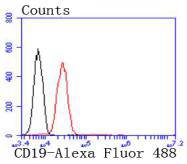
Fig2: Flow cytometric analysis of Jurkat cells with CD19 antibody at 1/50 dilution (red) compared with an unlabelled control (cells without incubation with primary antibody; black). Alexa Fluor 488-conjugated goat anti rabbit IgG was used as the secondary antibody| Product Name | CD19 [JF099-9] |
|---|---|
| Antibody Type | Primary Antibodies |
| Antigen Alias | antibody deficiency due to defect in CD19, included antibody AW495831 antibody B lymphocyte antigen CD19 antibody B lymphocyte surface antigen B4 antibody B-lymphocyte antigen CD19 antibody B-lymphocyte surface antigen B4 antibody B4 antibody CD19 antibody CD19 antigen antibody CD19 molecule antibody Cd19 protein antibody CD19_HUMAN antibody CVID3 antibody Differentiation antigen CD19 antibody Leu 12 antibody Leu-12 antibody Leu12 antibody MGC109570 antibody MGC12802 antibody T-cell surface antigen Leu-12 antibody |
| Product description | CD19 is a transmembrane glycoprotein that contains two extracellular immunoglobulin-like domains. CD19 is selectively expressed on the cell surface of B-lymphocytes, where it activates intracellular signaling cascades involving both Ras and phosphatidylinositol 3-kinase pathways. Activation of CD19 results in cross-linking of the membrane protein immunoglobulin chains and the subsequent association with Src family protein tyrosine kinases (PTK). Expression of CD19 is continuous throughout B-cell development and through terminal differentiation of B-cells into plasma cells. CD19 forms functional complexes with B-lymphocyte surface proteins, including integrin b1, CD21 and CD81, which are involved in regulating B-cell development. |
| Immunogen | recombinant protein |
| Clonality | Monoclonal |
|---|---|
| Isotype | IgG |
| Host Species | Recombinant rabbit |
| Tested Applications | WBFC |
| WB:1:1,000-1:5,000 FC:1:10-1:100 |
|
| Species Reactivity | HumanMouseRat |
| Concentration | 1mg/ml |
| Alternative Names | antibody
deficiency due to defect in CD19 included antibody AW495831 antibody B lymphocyte antigen CD19 antibody B lymphocyte surface antigen B4 antibody B-lymphocyte antigen CD19 antibody B-lymphocyte surface antigen B4 antibody B4 antibody CD19 antibody CD19 antigen antibody CD19 molecule antibody Cd19 protein antibody CD19_HUMAN antibody CVID3 antibody Differentiation antigen CD19 antibody Leu 12 antibody Leu-12 antibody Leu12 antibody MGC109570 antibody MGC12802 antibody T-cell surface antigen Leu-12 antibody |
|---|---|
| Molecular Weight(MW) | 75-100 kDa |
| Cellular Localization | Membrane. |
Application
Fig1: Western blot analysis of CD19 on different lysates using anti-CD19 antibody at 1/1,000 dilution. Positive control: Lane 1: Rat brain Lane 2: 293
Application
Fig2: Flow cytometric analysis of Jurkat cells with CD19 antibody at 1/50 dilution (red) compared with an unlabelled control (cells without incubation with primary antibody; black). Alexa Fluor 488-conjugated goat anti rabbit IgG was used as the secondary antibody| Positive Control | 293, rat brain tissue. |
|---|---|
| Application Notes | WB:1:1,000-1:5,000 FC:1:10-1:100 |
| Form | Liquid |
|---|---|
| Storage Instructions | Store at +4℃ after thawing. Aliquot store at -20℃ or -80℃. Avoid repeated freeze / thaw cycles. |
| Storage Buffer | 1*TBS (pH7.4), 1%BSA, 40%Glycerol. Preservative: 0.05% Sodium Azide. |


