Application
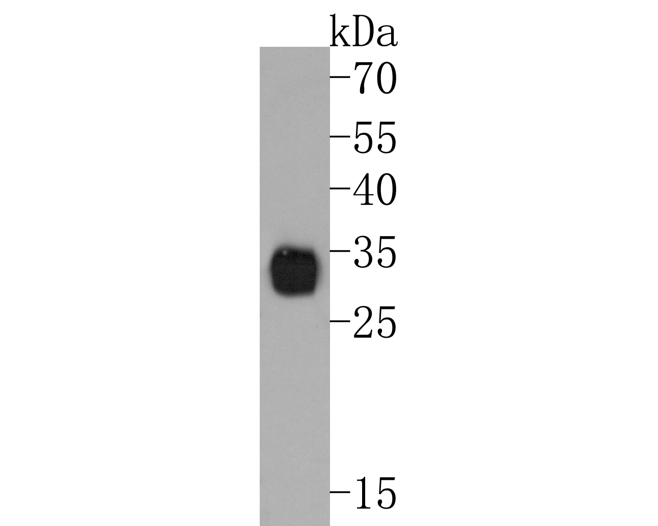
Fig1: Western blot analysis of Neutrophil Elastase on HL-60 cell lysates. Proteins were transferred to a PVDF membrane and blocked with 5% BSA in PBS for 1 hour at room temperature. The primary antibody (1/500) was used in 5% BSA at room temperature for 2 hours. Goat Anti-Rabbit IgG - HRP Secondary Antibody at 1:200,000 dilution was used for 1 hour at room temperature.Application
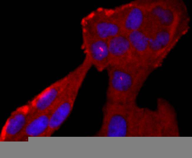
Fig2: ICC staining Neutrophil Elastase in Hela cells (red). The nuclear counter stain is DAPI (blue). Cells were fixed in paraformaldehyde, permeabilised with 0.25% Triton X100/PBS.Application
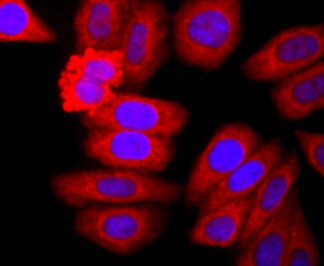
Fig3: ICC staining Neutrophil Elastase in HepG2 cells (red). The nuclear counter stain is DAPI (blue). Cells were fixed in paraformaldehyde, permeabilised with 0.25% Triton X100/PBS.Application
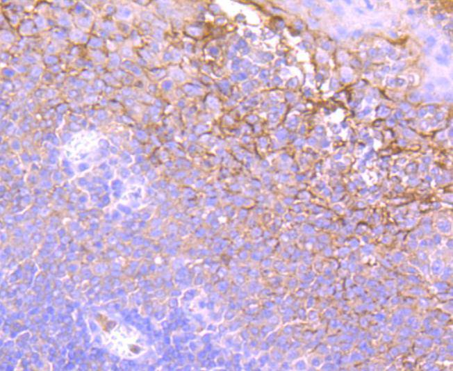
Fig4: Immunohistochemical analysis of paraffin-embedded human tonsil tissue using anti-Neutrophil Elastase antibody. Counter stained with hematoxylin.Application
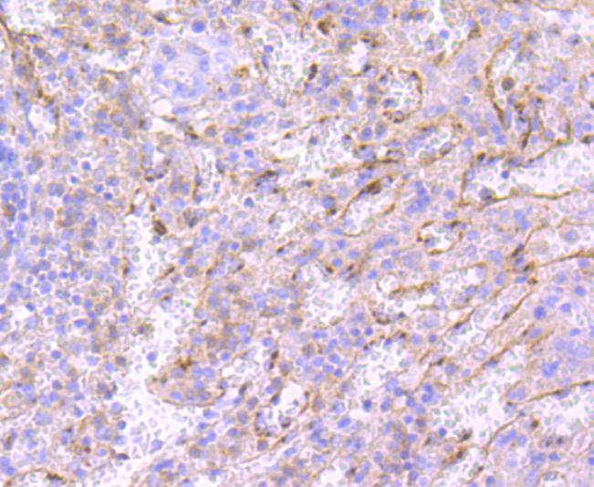
Fig5: Immunohistochemical analysis of paraffin-embedded human spleen tissue using anti-Neutrophil Elastase antibody. Counter stained with hematoxylin.Application
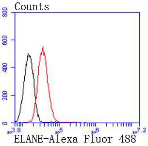
Fig6: Flow cytometric analysis of HepG2 cells with Neutrophil Elastase antibody at 1/50 dilution (red) compared with an unlabelled control (cells without incubation with primary antibody; black). Alexa Fluor 488-conjugated goat anti rabbit IgG was used as the secondary antibody| Product Name | Neutrophil Elastase Recombinant Rabbit Monoclonal Antibody |
|---|---|
| Antibody Type | Primary Antibodies |
| Product description | Neutrophil Elastase (NE) is a serine protease that is expressed in bone marrow precursor cells, stored in peripheral blood granulocytes, and implicated in the progression of a variety of inflammatory diseases, including idiopathic pulmonary fibrosis, rheumatoid arthritis, adult respiratory distress syndrome and cystic fibrosis. In neutrophils, Neutrophil Elastase contributes largely to the proteolysis of phagocytosed proteins, the migration of neutrophils and the remodeling of tissues following injury. Neutrophil Elastase, which is also designated medullasin, is secreted into the extracellular matrix, where it is then capable of destroying connective tissue proteins, including elastin, proteoglycans and Type IV Collagens. Neutrophil Elastase also mediates proteolysis by cleaving proteins that are associated with the complement system, such as antithrombin and Fibrinogen. Additionally, Neutrophil Elastase functions as a potent platelet agonist, where it potentiates the aggregation, secretion and mobilization of calcium in response to cathepsin G binding to platelet surface receptors. |
| Immunogen | Recombinant protein within Human Neutrophil Elastase aa 1-140 / 268. |
| Clonality | monoclonal |
|---|---|
| Isotype | IgG |
| Host Species | Recombinant rabbit |
| Tested Applications | FCICC/IFIHCWB |
| WB:1:500-1:2000 ICC:1:50 IHC:1:50-1:200 FC:1:10-1:50 |
|
| Species Reactivity | HumanMouse |
| Concentration | 1mg/ |
| Purification | Protein A |
| Alternative Names | Bone marrow serine protease antibody ELA2 antibody ELANE antibody Elastase 2 antibody Elastase 2 neutrophil antibody Elastase neutrophil expressed antibody Elastase-2 antibody ELNE_HUMAN antibody GE antibody Granulocyte derived elastase antibody HLE antibody HNE antibody Human leukocyte elastase antibody Leukocyte elastase antibody Medullasin antibody NE antibody Neutrophil elastase antibody PMN E antibody PMN elastase antibody Polymorphonuclear elastase antibody SCN1 antibody |
|---|---|
| Molecular Weight(MW) | 29kDa |
| Cellular Localization | Cell surface, cytoplasm, extracellular region, secretory granule. |
| SwissProt ID | P08246 |
|---|
Application
Fig1: Western blot analysis of Neutrophil Elastase on HL-60 cell lysates. Proteins were transferred to a PVDF membrane and blocked with 5% BSA in PBS for 1 hour at room temperature. The primary antibody (1/500) was used in 5% BSA at room temperature for 2 hours. Goat Anti-Rabbit IgG - HRP Secondary Antibody at 1:200,000 dilution was used for 1 hour at room temperature.
Application
Fig2: ICC staining Neutrophil Elastase in Hela cells (red). The nuclear counter stain is DAPI (blue). Cells were fixed in paraformaldehyde, permeabilised with 0.25% Triton X100/PBS.
Application
Fig3: ICC staining Neutrophil Elastase in HepG2 cells (red). The nuclear counter stain is DAPI (blue). Cells were fixed in paraformaldehyde, permeabilised with 0.25% Triton X100/PBS.
Application
Fig4: Immunohistochemical analysis of paraffin-embedded human tonsil tissue using anti-Neutrophil Elastase antibody. Counter stained with hematoxylin.
Application
Fig5: Immunohistochemical analysis of paraffin-embedded human spleen tissue using anti-Neutrophil Elastase antibody. Counter stained with hematoxylin.
Application
Fig6: Flow cytometric analysis of HepG2 cells with Neutrophil Elastase antibody at 1/50 dilution (red) compared with an unlabelled control (cells without incubation with primary antibody; black). Alexa Fluor 488-conjugated goat anti rabbit IgG was used as the secondary antibody| Positive Control | A549, Hela, HepG2, human tonsil tissue, human spleen tissue. |
|---|---|
| Application Notes | WB:1:500-1:2000 ICC:1:50 IHC:1:50-1:200 FC:1:10-1:50 |
| Form | Liquid |
|---|---|
| Storage Instructions | Store at +4℃ after thawing. Aliquot store at -20℃ or -80℃. Avoid repeated freeze / thaw cycles. |
| Storage Buffer | 1*TBS (pH7.4), 1%BSA, 40%Glycerol. Preservative: 0.05% Sodium Azide. |


