WB
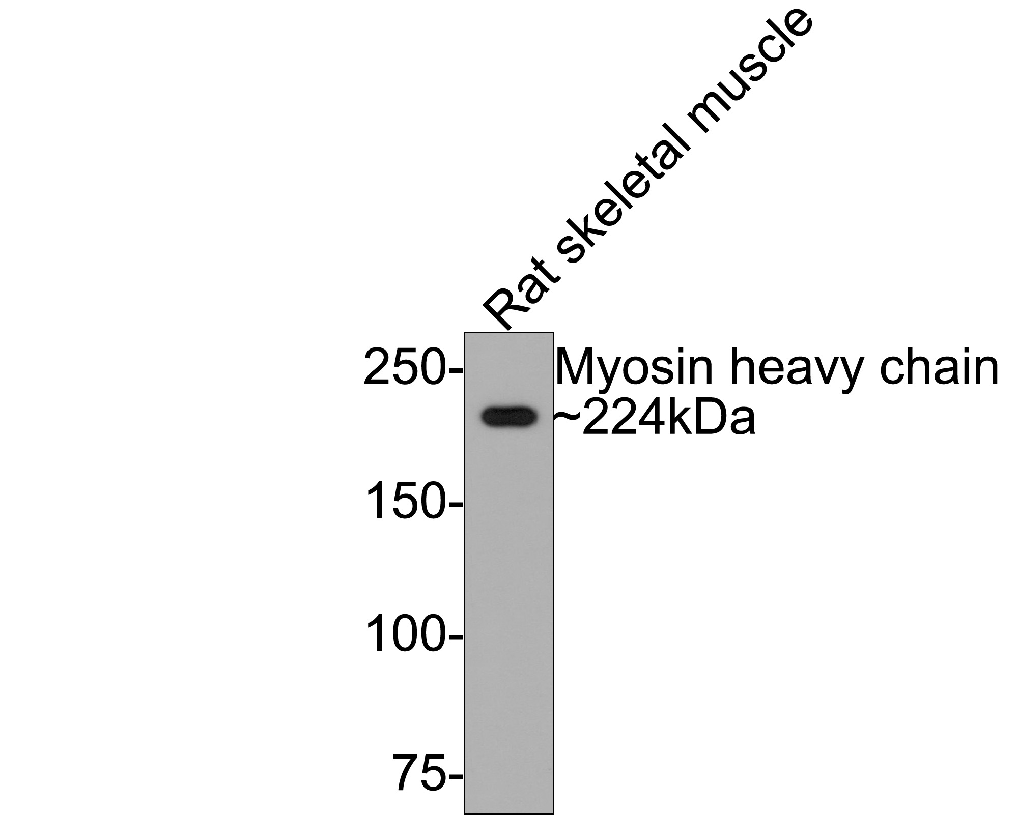
Western blot analysis of Myosin heavy chain on rat skeletal muscle tissue lysates with Rabbit anti-Myosin heavy chain antibody at 1/500 dilution. Lysates/proteins at 20 µg/Lane. Exposure time: 2 minutes; 6% SDS-PAGE gel. Proteins were transferred to a PVDF membrane and blocked with 5% NFDM/TBST for 1 hour at room temperature. The primary antibody at 1/500 dilution was used in 5% NFDM/TBST at room temperature for 2 hours. Goat Anti-Rabbit IgG - HRP Secondary Antibody at 1:300,000 dilution was used for 1 hour at room temperature.IHC
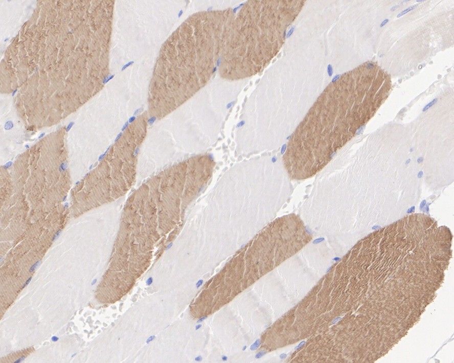
Immunohistochemical analysis of paraffin-embedded human skeletal muscle tissue with Rabbit anti-Myosin heavy chain antibody at 1/2,000 dilution. The section was pre-treated using heat mediated antigen retrieval with Tris-EDTA buffer (pH 9.0) for 20 minutes. The tissues were blocked in 1% BSA for 20 minutes at room temperature, washed with ddH2O and PBS, and then probed with the primary antibody at 1/2,000 dilution for 1 hour at room temperature. The detection was performed using an HRP conjugated compact polymer system. DAB was used as the chromogen. Tissues were counterstained with hematoxylin and mounted with DPX.IHC
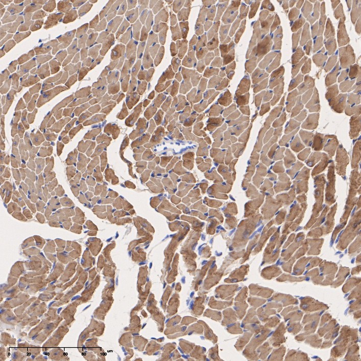
Immunohistochemical analysis of paraffin-embedded mouse heart tissue with Rabbit anti-Myosin heavy chain antibody at 1/400 dilution. The section was pre-treated using heat mediated antigen retrieval with Tris-EDTA buffer (pH 9.0) for 20 minutes. The tissues were blocked in 1% BSA for 20 minutes at room temperature, washed with ddH2O and PBS, and then probed with the primary antibody at 1/400 dilution for 1 hour at room temperature. The detection was performed using an HRP conjugated compact polymer system. DAB was used as the chromogen. Tissues were counterstained with hematoxylin and mounted with DPX.IHC
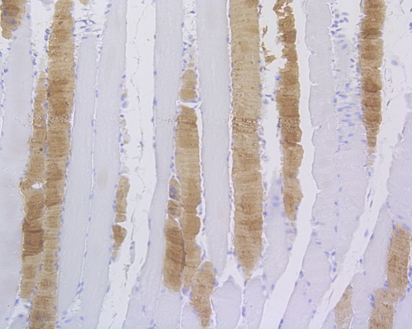
Immunohistochemical analysis of paraffin-embedded rat skeletal muscle tissue using anti-Myosin heavy chain antibody. The section was pre-treated using heat mediated antigen retrieval with Tris-EDTA buffer (pH 8.0-8.4) for 20 minutes.The tissues were blocked in 5% BSA for 30 minutes at room temperature, washed with ddH2O and PBS, and then probed with the primary antibody (1/400) for 30 minutes at room temperature. The detection was performed using an HRP conjugated compact polymer system. DAB was used as the chromogen. Tissues were counterstained with hematoxylin and mounted with DPX.IF-P
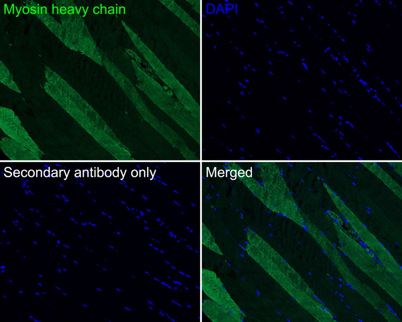
Immunofluorescence analysis of paraffin-embedded human striated muscle tissue labeling Myosin heavy chain with Rabbit anti-Myosin heavy chain antibody at 1/100 dilution. The section was pre-treated using heat mediated antigen retrieval with Tris-EDTA buffer (pH 9.0) for 20 minutes. The tissues were blocked in 10% negative goat serum for 1 hour at room temperature, washed with PBS, and then probed with the primary antibody (green) at 1/100 dilution overnight at 4 ℃, washed with PBS. Goat Anti-Rabbit IgG H&L (488) was used as the secondary antibody at 1/1,000 dilution. Nuclei were counterstained with DAPI (blue).IF-P
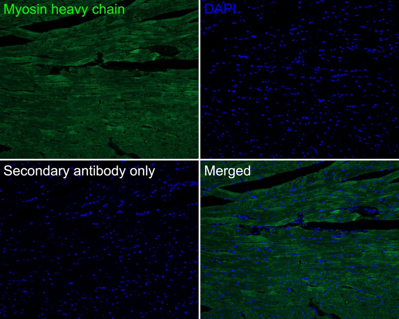
Immunofluorescence analysis of paraffin-embedded mouse heart tissue labeling Myosin heavy chain with Rabbit anti-Myosin heavy chain antibody at 1/100 dilution. The section was pre-treated using heat mediated antigen retrieval with Tris-EDTA buffer (pH 9.0) for 20 minutes. The tissues were blocked in 10% negative goat serum for 1 hour at room temperature, washed with PBS, and then probed with the primary antibody (green) at 1/100 dilution overnight at 4 ℃, washed with PBS. Goat Anti-Rabbit IgG H&L (488) was used as the secondary antibody at 1/1,000 dilution. Nuclei were counterstained with DAPI (blue).IF-P
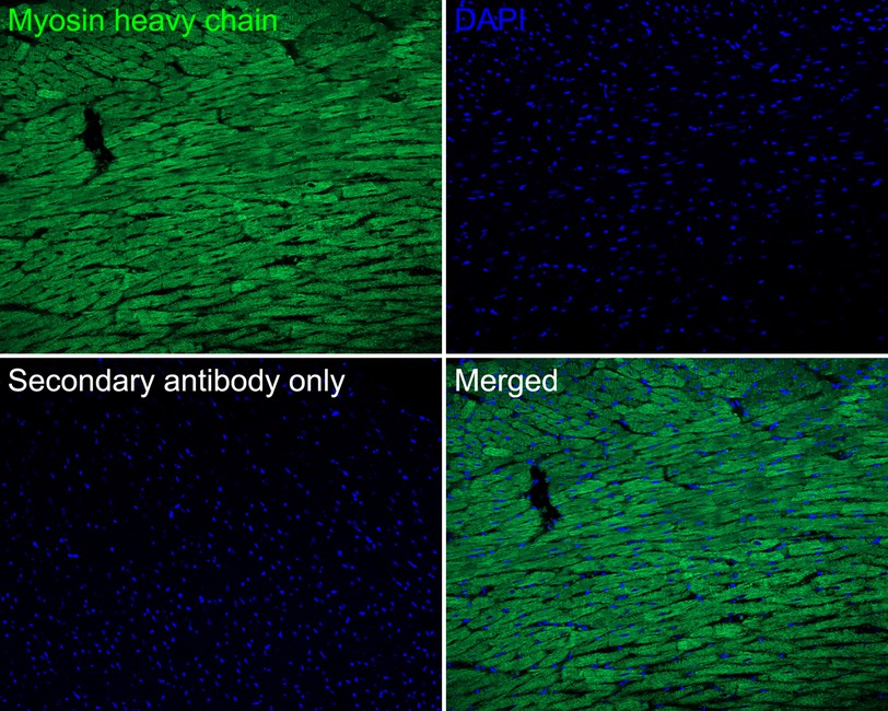
Immunofluorescence analysis of paraffin-embedded rat heart tissue labeling Myosin heavy chain with Rabbit anti-Myosin heavy chain antibody at 1/100 dilution. The section was pre-treated using heat mediated antigen retrieval with Tris-EDTA buffer (pH 9.0) for 20 minutes. The tissues were blocked in 10% negative goat serum for 1 hour at room temperature, washed with PBS, and then probed with the primary antibody (green) at 1/100 dilution overnight at 4 ℃, washed with PBS. Goat Anti-Rabbit IgG H&L (488) was used as the secondary antibody at 1/1,000 dilution. Nuclei were counterstained with DAPI (blue).| Product Name | Myosin heavy chain Recombinant Rabbit Monoclonal Antibody |
|---|---|
| Antibody Type | Primary Antibodies |
| Product description | Myosin is a highly conserved, ubiquitously expressed protein that interacts with Actin to generate the force for cellular movements. Conventional myosins are hexameric proteins consisting of two heavy chain subunits, a pair of non-phosphorylatable light chain subunits and a pair of phosphorylatable light chain subunits. Three general classes of myosin have been cloned: smooth muscle myosins, striated muscle myosins and non-muscle myosins. Contractile activity in smooth muscle is regulated by the calcium/calmodulin-dependent phosphorylation of myosin light chain by myosin light chain kinase. Myosin heavy chains are encoded by the MYH gene family and have Actin-activated ATPase activity which generates the motor function of myosin. Myosin heavy chains, which were initially isolated from a human fetal skeletal muscle, are the major determinant in the speed of contraction of skeletal muscle. Various isoforms of myosin heavy chain are differentially expressed depending on the functional activity of the muscle. |
| Immunogen | Recombinant protein within Human Myosin heavy chain aa 1220-1420 / 1939. |
| Clonality | Monoclonal |
|---|---|
| Isotype | IgG |
| Host Species | Recombinant rabbit |
| Tested Applications | IF-PIHCWB |
| WB:1:500-1:2000 IHC:1:100-1:500 IF-P:1:100 |
|
| Species Reactivity | HumanMouseRat |
| Concentration | 1mg/ml |
| Purification | Protein A |
| Alternative Names | cardiac muscle alpha isoform antibody MYH6 antibody MYH6_HUMAN antibody MYH7 antibody MYHC antibody MyHC-alpha antibody MyHC-beta antibody MYHCA antibody MYHCB antibody Myosin heavy chain 6 antibody Myosin heavy chain antibody Myosin heavy chain cardiac muscle alpha isoform antibody Myosin heavy chain cardiac muscle beta isoform antibody Myosin-6 antibody |
|---|---|
| Molecular Weight(MW) | 224 kDa |
| Cellular Localization | Cytoplasm, myofibril. |
WB
Western blot analysis of Myosin heavy chain on rat skeletal muscle tissue lysates with Rabbit anti-Myosin heavy chain antibody at 1/500 dilution. Lysates/proteins at 20 µg/Lane. Exposure time: 2 minutes; 6% SDS-PAGE gel. Proteins were transferred to a PVDF membrane and blocked with 5% NFDM/TBST for 1 hour at room temperature. The primary antibody at 1/500 dilution was used in 5% NFDM/TBST at room temperature for 2 hours. Goat Anti-Rabbit IgG - HRP Secondary Antibody at 1:300,000 dilution was used for 1 hour at room temperature.
IHC
Immunohistochemical analysis of paraffin-embedded human skeletal muscle tissue with Rabbit anti-Myosin heavy chain antibody at 1/2,000 dilution. The section was pre-treated using heat mediated antigen retrieval with Tris-EDTA buffer (pH 9.0) for 20 minutes. The tissues were blocked in 1% BSA for 20 minutes at room temperature, washed with ddH2O and PBS, and then probed with the primary antibody at 1/2,000 dilution for 1 hour at room temperature. The detection was performed using an HRP conjugated compact polymer system. DAB was used as the chromogen. Tissues were counterstained with hematoxylin and mounted with DPX.
IHC
Immunohistochemical analysis of paraffin-embedded mouse heart tissue with Rabbit anti-Myosin heavy chain antibody at 1/400 dilution. The section was pre-treated using heat mediated antigen retrieval with Tris-EDTA buffer (pH 9.0) for 20 minutes. The tissues were blocked in 1% BSA for 20 minutes at room temperature, washed with ddH2O and PBS, and then probed with the primary antibody at 1/400 dilution for 1 hour at room temperature. The detection was performed using an HRP conjugated compact polymer system. DAB was used as the chromogen. Tissues were counterstained with hematoxylin and mounted with DPX.
IHC
Immunohistochemical analysis of paraffin-embedded rat skeletal muscle tissue using anti-Myosin heavy chain antibody. The section was pre-treated using heat mediated antigen retrieval with Tris-EDTA buffer (pH 8.0-8.4) for 20 minutes.The tissues were blocked in 5% BSA for 30 minutes at room temperature, washed with ddH2O and PBS, and then probed with the primary antibody (1/400) for 30 minutes at room temperature. The detection was performed using an HRP conjugated compact polymer system. DAB was used as the chromogen. Tissues were counterstained with hematoxylin and mounted with DPX.
IF-P
Immunofluorescence analysis of paraffin-embedded human striated muscle tissue labeling Myosin heavy chain with Rabbit anti-Myosin heavy chain antibody at 1/100 dilution. The section was pre-treated using heat mediated antigen retrieval with Tris-EDTA buffer (pH 9.0) for 20 minutes. The tissues were blocked in 10% negative goat serum for 1 hour at room temperature, washed with PBS, and then probed with the primary antibody (green) at 1/100 dilution overnight at 4 ℃, washed with PBS. Goat Anti-Rabbit IgG H&L (488) was used as the secondary antibody at 1/1,000 dilution. Nuclei were counterstained with DAPI (blue).
IF-P
Immunofluorescence analysis of paraffin-embedded mouse heart tissue labeling Myosin heavy chain with Rabbit anti-Myosin heavy chain antibody at 1/100 dilution. The section was pre-treated using heat mediated antigen retrieval with Tris-EDTA buffer (pH 9.0) for 20 minutes. The tissues were blocked in 10% negative goat serum for 1 hour at room temperature, washed with PBS, and then probed with the primary antibody (green) at 1/100 dilution overnight at 4 ℃, washed with PBS. Goat Anti-Rabbit IgG H&L (488) was used as the secondary antibody at 1/1,000 dilution. Nuclei were counterstained with DAPI (blue).
IF-P
Immunofluorescence analysis of paraffin-embedded rat heart tissue labeling Myosin heavy chain with Rabbit anti-Myosin heavy chain antibody at 1/100 dilution. The section was pre-treated using heat mediated antigen retrieval with Tris-EDTA buffer (pH 9.0) for 20 minutes. The tissues were blocked in 10% negative goat serum for 1 hour at room temperature, washed with PBS, and then probed with the primary antibody (green) at 1/100 dilution overnight at 4 ℃, washed with PBS. Goat Anti-Rabbit IgG H&L (488) was used as the secondary antibody at 1/1,000 dilution. Nuclei were counterstained with DAPI (blue).| Application Notes | WB:1:500-1:2000 IHC:1:100-1:500 IF-P:1:100 |
|---|
| Form | Liquid |
|---|---|
| Storage Instructions | Store at +4℃ after thawing. Aliquot store at -20℃ or -80℃. Avoid repeated freeze / thaw cycles. |
| Storage Buffer | 1*TBS (pH7.4), 1%BSA, 40%Glycerol. Preservative: 0.05% Sodium Azide. |


