WB
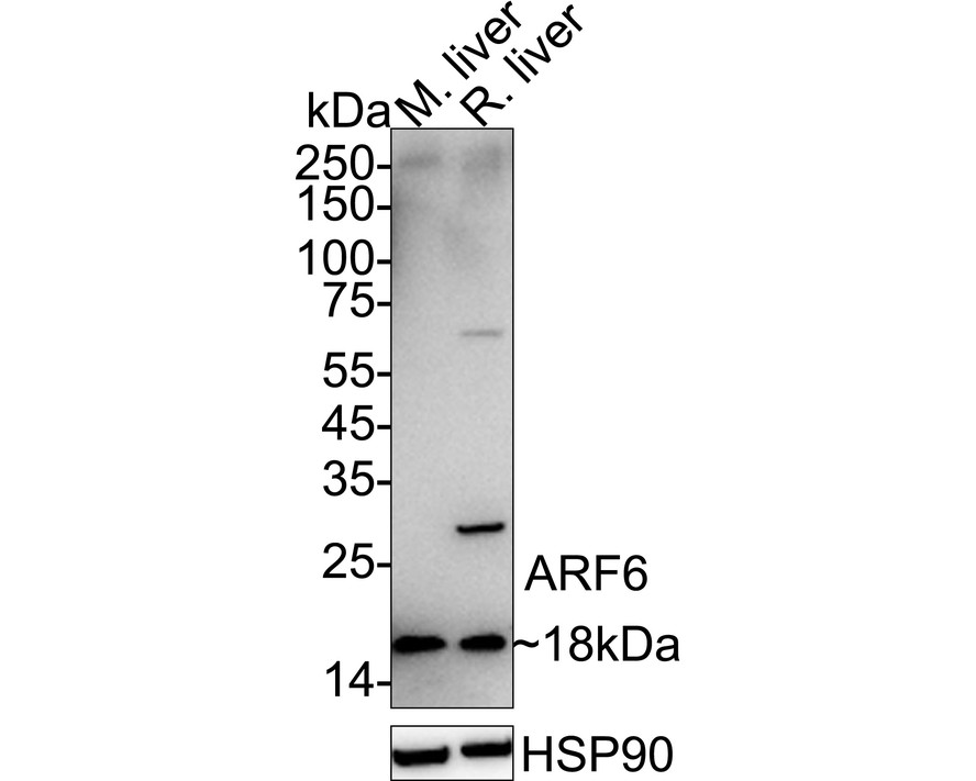
Western blot analysis of ARF6 on different lysates with Rabbit anti-ARF6 antibody at 1/1,000 dilution. Lane 1: Mouse liver tissue lysate, Lane 2: Rat liver tissue lysate, Lysates/proteins at 40 µg/Lane. Exposure time: 3 minutes; 4-20% SDS-PAGE gel. Proteins were transferred to a PVDF membrane and blocked with 5% NFDM/TBST for 1 hour at room temperature. The primary antibody at 1/1,000 dilution was used in 5% NFDM/TBST at 4℃ overnight. Goat Anti-Rabbit IgG - HRP Secondary Antibody at 1/50,000 dilution was used for 1 hour at room temperature.WB
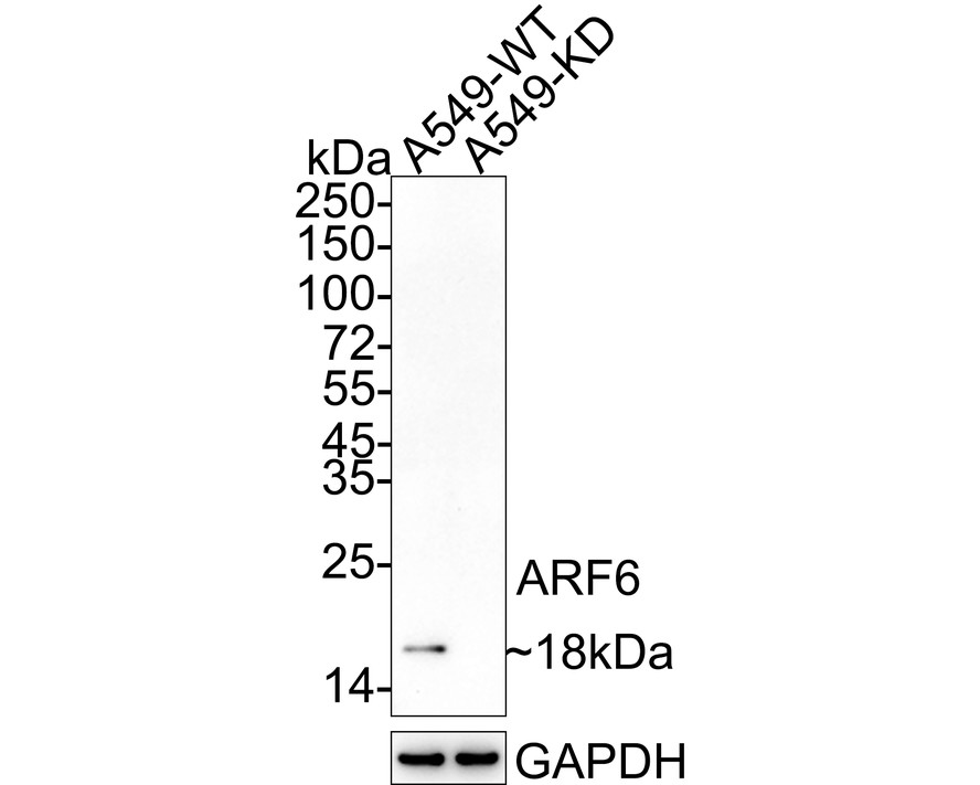
Western blot analysis of ARF6 on different lysates with Rabbit anti-ARF6 antibody at 1/1,000 dilution. Lane 1: A549-WT cell lysate, Lane 2: A549-KD ARF6 cell lysate, Lysates/proteins at 10 µg/Lane. Exposure time: 3 minutes; 4-20% SDS-PAGE gel. Proteins were transferred to a PVDF membrane and blocked with 5% NFDM/TBST for 1 hour at room temperature. The primary antibody at 1/1,000 dilution was used in 5% NFDM/TBST at 4℃ overnight. Goat Anti-Rabbit IgG - HRP Secondary Antibody at 1/50,000 dilution was used for 1 hour at room temperature.IHC
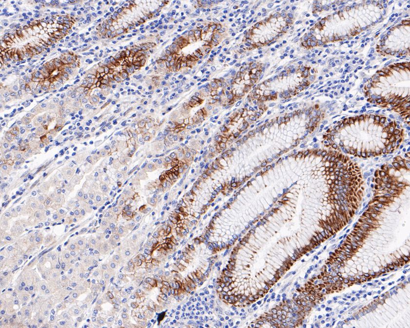
Immunohistochemical analysis of paraffin-embedded human stomach tissue using anti-ARF6 antibody. The section was pre-treated using heat mediated antigen retrieval with Tris-EDTA buffer (pH 8.0-8.4) for 20 minutes.The tissues were blocked in 5% BSA for 30 minutes at room temperature, washed with ddH2O and PBS, and then probed with the primary antibody (1/200) for 30 minutes at room temperature. The detection was performed using an HRP conjugated compact polymer system. DAB was used as the chromogen. Tissues were counterstained with hematoxylin and mounted with DPX.ICC/IF
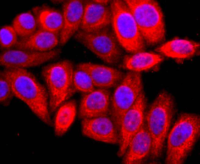
ICC staining of ARF6 in HepG2 cells (red). Formalin fixed cells were permeabilized with 0.1% Triton X-100 in TBS for 10 minutes at room temperature and blocked with 1% Blocker BSA for 15 minutes at room temperature. Cells were probed with the primary antibody (1/50) for 1 hour at room temperature, washed with PBS. Alexa Fluor®594 Goat anti-Rabbit IgG was used as the secondary antibody at 1/1,000 dilution. The nuclear counter stain is DAPI (blue).IF-F
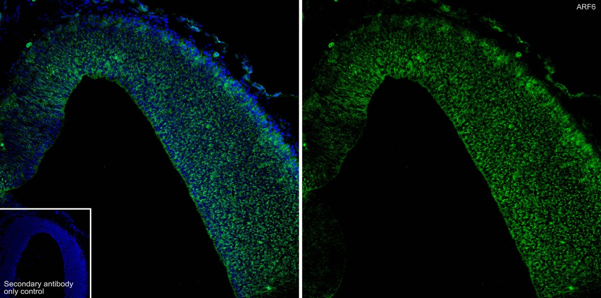
Immunofluorescence analysis of frozen mouse embryonic brain tissue with Rabbit anti-ARF6 antibody at 1/500 dilution. The section was not undergone antigen retrieval. The tissues were blocked in 10% negative goat serum for 1 hour at room temperature, washed with PBS, and then probed with the primary antibody (green) at 1/500 dilution overnight at 4 ℃, washed with PBS. Goat Anti-Rabbit IgG H&L (488) was used as the secondary antibody at 1/1,000 dilution. Nuclei were counterstained with DAPI (blue).IF-P
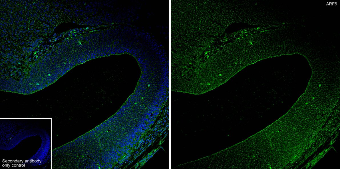
Immunofluorescence analysis of paraffin-embedded mouse embryonic brain tissue labeling ARF6 with Rabbit anti-ARF6 antibody at 1/200 dilution. The section was pre-treated using heat mediated antigen retrieval with Tris-EDTA buffer (pH 9.0) for 20 minutes. The tissues were blocked in 10% negative goat serum for 1 hour at room temperature, washed with PBS, and then probed with the primary antibody (green) at 1/200 dilution overnight at 4 ℃, washed with PBS. Goat Anti-Rabbit IgG H&L (488) was used as the secondary antibody at 1/1,000 dilution. Nuclei were counterstained with DAPI (blue).| Product Name | ARF6 Recombinant Rabbit Monoclonal Antibody |
|---|---|
| Antibody Type | Primary Antibodies |
| Product description | The ADP-ribosylation factor (ARF) protein family are structurally and functionally conserved members of the Ras superfamily of regulatory GTP-binding proteins. ARFs influence vesicle trafficking and signal transduction in eukaryotic cells. ARF-dependent regulatory mechanisms include the coordination of spectrin interactions with golgi membranes and the association of Actin to the Golgi via rho family-dependent G-protein localization (Rac, CDC42) and WASP/Arp2/3 complexes. Additionally, ARFs play a central role in maintenance of organelle integrity, assembly of coat proteins and activation of phospholipase D. The ARF proteins are categorized as class I (ARF1, ARF2 and ARF3), class II (ARF4 and ARF5) and class III (ARF6); members of each class share a common gene organization. The human ARF6 gene contains five exons and four introns, and encodes a 175 amino acid protein. |
| Immunogen | Synthetic peptide within Human ARF6 aa 133-165 / 175. |
| Clonality | Monoclonal |
|---|---|
| Isotype | IgG |
| Host Species | Recombinant rabbit |
| Tested Applications | ICC/IFIF-FIF-PIHCWB |
| WB:1:500-1:2000 IHC:1:50-1:200 ICC/IF:1:100-1:500 IF-F:1:500 IF-P:1:200 |
|
| Species Reactivity | HumanMouseRat |
| Concentration | 1mg/ml |
| Purification | Protein A |
| Alternative Names | ADP ribosylation factor 6 antibody ADP ribosylation factor protein 6 antibody ADP-ribosylation factor 6 antibody ARF6 antibody ARF6_HUMAN antibody DKFZp564M0264 antibody Small GTP binding protein antibody Small GTPase antibody |
|---|---|
| Molecular Weight(MW) | 20 kDa(Observed band size: 18 kDa) |
| Cellular Localization | Golgi apparatus, Cell membrane, Endosome membrane, Cell projection, Midbody, Cytoplasm, Cleavage furrow. |
WB
Western blot analysis of ARF6 on different lysates with Rabbit anti-ARF6 antibody at 1/1,000 dilution. Lane 1: Mouse liver tissue lysate, Lane 2: Rat liver tissue lysate, Lysates/proteins at 40 µg/Lane. Exposure time: 3 minutes; 4-20% SDS-PAGE gel. Proteins were transferred to a PVDF membrane and blocked with 5% NFDM/TBST for 1 hour at room temperature. The primary antibody at 1/1,000 dilution was used in 5% NFDM/TBST at 4℃ overnight. Goat Anti-Rabbit IgG - HRP Secondary Antibody at 1/50,000 dilution was used for 1 hour at room temperature.
WB
Western blot analysis of ARF6 on different lysates with Rabbit anti-ARF6 antibody at 1/1,000 dilution. Lane 1: A549-WT cell lysate, Lane 2: A549-KD ARF6 cell lysate, Lysates/proteins at 10 µg/Lane. Exposure time: 3 minutes; 4-20% SDS-PAGE gel. Proteins were transferred to a PVDF membrane and blocked with 5% NFDM/TBST for 1 hour at room temperature. The primary antibody at 1/1,000 dilution was used in 5% NFDM/TBST at 4℃ overnight. Goat Anti-Rabbit IgG - HRP Secondary Antibody at 1/50,000 dilution was used for 1 hour at room temperature.
IHC
Immunohistochemical analysis of paraffin-embedded human stomach tissue using anti-ARF6 antibody. The section was pre-treated using heat mediated antigen retrieval with Tris-EDTA buffer (pH 8.0-8.4) for 20 minutes.The tissues were blocked in 5% BSA for 30 minutes at room temperature, washed with ddH2O and PBS, and then probed with the primary antibody (1/200) for 30 minutes at room temperature. The detection was performed using an HRP conjugated compact polymer system. DAB was used as the chromogen. Tissues were counterstained with hematoxylin and mounted with DPX.
ICC/IF
ICC staining of ARF6 in HepG2 cells (red). Formalin fixed cells were permeabilized with 0.1% Triton X-100 in TBS for 10 minutes at room temperature and blocked with 1% Blocker BSA for 15 minutes at room temperature. Cells were probed with the primary antibody (1/50) for 1 hour at room temperature, washed with PBS. Alexa Fluor®594 Goat anti-Rabbit IgG was used as the secondary antibody at 1/1,000 dilution. The nuclear counter stain is DAPI (blue).
IF-F
Immunofluorescence analysis of frozen mouse embryonic brain tissue with Rabbit anti-ARF6 antibody at 1/500 dilution. The section was not undergone antigen retrieval. The tissues were blocked in 10% negative goat serum for 1 hour at room temperature, washed with PBS, and then probed with the primary antibody (green) at 1/500 dilution overnight at 4 ℃, washed with PBS. Goat Anti-Rabbit IgG H&L (488) was used as the secondary antibody at 1/1,000 dilution. Nuclei were counterstained with DAPI (blue).
IF-P
Immunofluorescence analysis of paraffin-embedded mouse embryonic brain tissue labeling ARF6 with Rabbit anti-ARF6 antibody at 1/200 dilution. The section was pre-treated using heat mediated antigen retrieval with Tris-EDTA buffer (pH 9.0) for 20 minutes. The tissues were blocked in 10% negative goat serum for 1 hour at room temperature, washed with PBS, and then probed with the primary antibody (green) at 1/200 dilution overnight at 4 ℃, washed with PBS. Goat Anti-Rabbit IgG H&L (488) was used as the secondary antibody at 1/1,000 dilution. Nuclei were counterstained with DAPI (blue).| Application Notes | WB:1:500-1:2000 IHC:1:50-1:200 ICC/IF:1:100-1:500 IF-F:1:500 IF-P:1:200 |
|---|
| Form | Liquid |
|---|---|
| Storage Instructions | Store at +4℃ after thawing. Aliquot store at -20℃ or -80℃. Avoid repeated freeze / thaw cycles. |
| Storage Buffer | 1*TBS (pH7.4), 1%BSA, 40%Glycerol. Preservative: 0.05% Sodium Azide. |


