WB
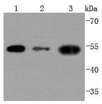
Western blot analysis of THY1 on different lysates using anti-THY1 antibody at 1/1,000 dilution. Positive control: Lane 1: Hela Lane 2: HepG2 Lane 3: HUVECICC/IF
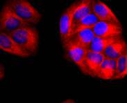
ICC staining THY1 in Hela cells (red). The nuclear counter stain is DAPI (blue). Cells were fixed in paraformaldehyde, permeabilised with 0.25% Triton X100/PBS.ICC/IF
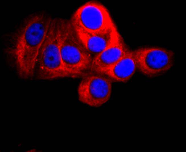
ICC staining THY1 in MCF-7 cells (red). The nuclear counter stain is DAPI (blue). Cells were fixed in paraformaldehyde, permeabilised with 0.25% Triton X100/PBS.ICC/IF
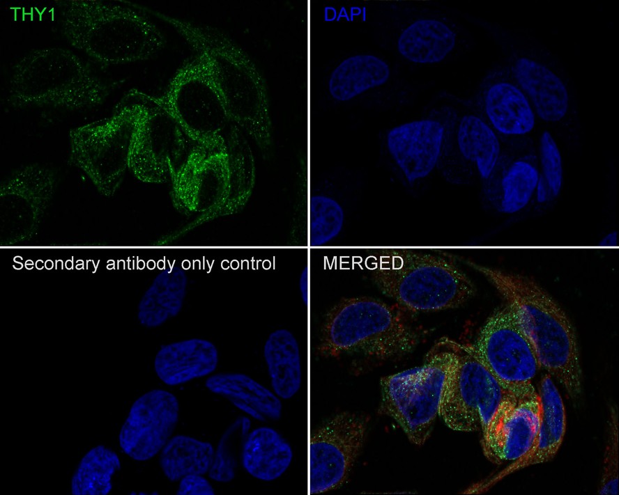
Immunocytochemistry analysis of HeLa cells labeling CD90 / THY1 with Rabbit anti-CD90 / THY1 antibody at 1/200 dilution. Cells were fixed in 4% paraformaldehyde for 10 minutes at 37 ℃, permeabilized with 0.05% Triton X-100 in PBS for 20 minutes, and then blocked with 2% negative goat serum for 30 minutes at room temperature. Cells were then incubated with Rabbit anti-CD90 / THY1 antibody at 1/200 dilution in 2% negative goat serum overnight at 4 ℃. Goat Anti-Rabbit IgG H&L (488) was used as the secondary antibody at 1/1,000 dilution. Nuclear DNA was labelled in blue with DAPI. Beta tubulin (red) was stained at 1/200 dilution overnight at +4℃. Goat Anti-Mouse IgG H&L (594) was used as the secondary antibody at 1/1,000 dilution.IHC
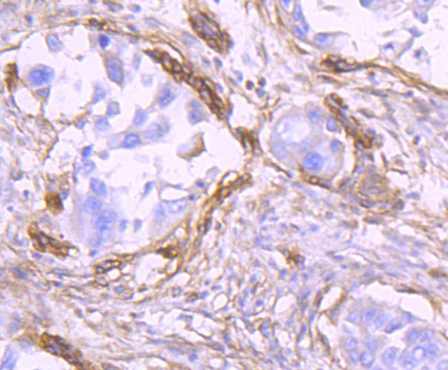
Immunohistochemical analysis of paraffin-embedded human lung cancer tissue using anti-THY1 antibody. Counter stained with hematoxylin.IHC
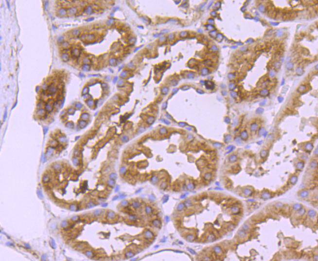
Immunohistochemical analysis of paraffin-embedded human kidney tissue using anti-THY1 antibody. Counter stained with hematoxylin.IHC
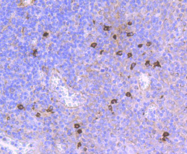
Immunohistochemical analysis of paraffin-embedded human tonsil tissue using anti-THY1 antibody. Counter stained with hematoxylin.IHC
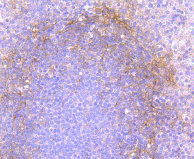
Immunohistochemical analysis of paraffin-embedded human spleen tissue using anti-THY1 antibody. Counter stained with hematoxylin.IHC
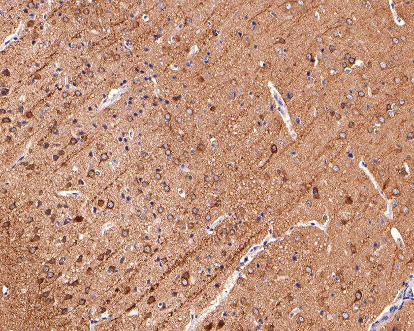
Immunohistochemical analysis of paraffin-embedded mouse brain tissue using anti-CD90 / THY1 antibody. The section was pre-treated using heat mediated antigen retrieval with Tris-EDTA buffer (pH 9.0) for 20 minutes.The tissues were blocked in 1% BSA for 30 minutes at room temperature, washed with ddH2O and PBS, and then probed with the primary antibody (1/400) for 30 minutes at room temperature. The detection was performed using an HRP conjugated compact polymer system. DAB was used as the chromogen. Tissues were counterstained with hematoxylin and mounted with DPX.IHC
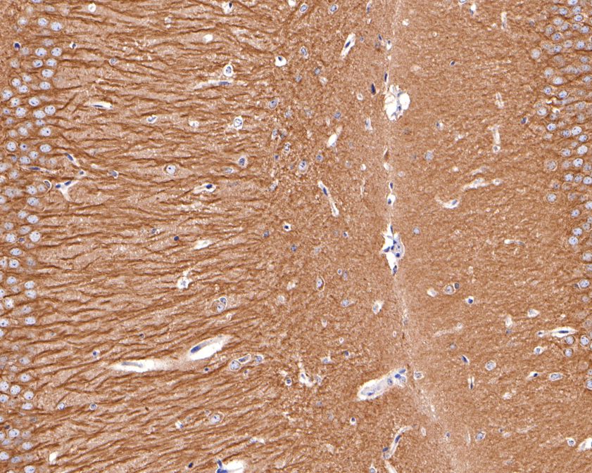
Immunohistochemical analysis of paraffin-embedded mouse hippocampus tissue using anti-CD90 / THY1 antibody. The section was pre-treated using heat mediated antigen retrieval with Tris-EDTA buffer (pH 9.0) for 20 minutes.The tissues were blocked in 1% BSA for 30 minutes at room temperature, washed with ddH2O and PBS, and then probed with the primary antibody (1/400) for 30 minutes at room temperature. The detection was performed using an HRP conjugated compact polymer system. DAB was used as the chromogen. Tissues were counterstained with hematoxylin and mounted with DPX.IF-F
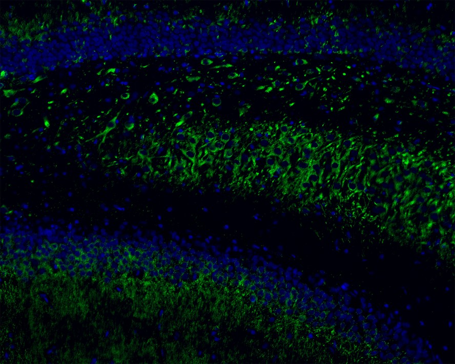
Immunofluorescence analysis of frozen mouse hippocampus tissue labeling CD90 / THY1 with Rabbit anti-CD90 / THY1 antibody. The tissues were blocked in 3% BSA for 30 minutes at room temperature, washed with PBS, and then probed with the primary antibody (green) at 1/100 dilution overnight at 4℃, washed with PBS. Goat Anti-Rabbit IgG H&L (488) was used as the secondary antibody at 1/200 dilution. Nuclei were counterstained with DAPI (blue). Image acquisition was performed with KFBIO KF-FL-400 Scanner.IF-P
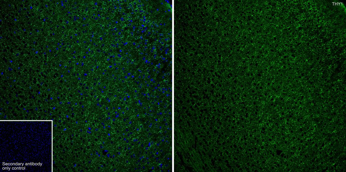
Immunofluorescence analysis of paraffin-embedded mouse brain tissue labeling CD90 / THY1 with Rabbit anti-CD90 / THY1 antibody at 1/400 dilution. The section was pre-treated using heat mediated antigen retrieval with Tris-EDTA buffer (pH 9.0) for 20 minutes. The tissues were blocked in 10% negative goat serum for 1 hour at room temperature, washed with PBS, and then probed with the primary antibody green) at 1/400 dilution overnight at 4 ℃, washed with PBS. Goat Anti-Rabbit IgG H&L (488) was used as the secondary antibody at 1/1,000 dilution. Nuclei were counterstained with DAPI (blue).| Product Name | CD90 / THY1 Recombinant Rabbit Monoclonal Antibody |
|---|---|
| Antibody Type | Primary Antibodies |
| Product description | Over 100 cell surface markers have been identified through the use of monoclonal antibodies. Many of these markers have proven useful in identifying specific subpopulations of cells within mixed colonies. Accordingly, these molecules have been assigned a "cluster of differentiation" (CD) designation. One such marker, designated Thy-1 (also referred to as CDw90), is a phosphatidyl-anchored cell surface glycoprotein which, when coexpressed with CD34 on cells from normal human bone marrow, identifies a subpopulation that includes putative hematopoietic, pleuripotent stem cells. Thy-1+ cells from bone marrow have been implicated in syngeneic graft versus host disease and may serve to regulate autoreactivity after bone marrow transplant. |
| Immunogen | Synthetic peptide within Human THY1 aa 75-120 / 161. |
| Clonality | Monoclonal |
|---|---|
| Isotype | IgG |
| Host Species | Recombinant rabbit |
| Tested Applications | ICC/IFIF-FIF-PIHCWB |
| WB:1:500-1:2000 ICC/IF:1:50-1:200 IHC:1:50-1:200 IF-F:1:100 IF-P:1:400 |
|
| Species Reactivity | HumanMouseRat |
| Concentration | 1mg/ml |
| Purification | Protein A |
| Alternative Names | CD7 antibody
CD90 antibody
CD90 antigen antibody
CDw90 antibody
FLJ33325 antibody
MGC128895 antibody
T25 antibody
Theta antigen antibody
Thy 1 antibody
Thy 1 cell surface antigen antibody
Thy 1 membrane glycoprotein antibody
Thy 1 T cell antigen antibody
Thy 1.2 antibody
Thy-1 antigen antibody
Thy-1 membrane glycoprotein antibody
Thy1 antibody
Thy1 antigen antibody
Thy1 T cell antigen antibody
Thy1.1 antibody
Thy1.2 antibody
THY1_HUMAN antibody
Thymus cell antigen 1 theta antibody |
|---|---|
| Molecular Weight(MW) | 45 kDa |
| Cellular Localization | Cell membrane. |
| SwissProt ID | P04216 |
|---|
WB
Western blot analysis of THY1 on different lysates using anti-THY1 antibody at 1/1,000 dilution. Positive control: Lane 1: Hela Lane 2: HepG2 Lane 3: HUVEC
ICC/IF
ICC staining THY1 in Hela cells (red). The nuclear counter stain is DAPI (blue). Cells were fixed in paraformaldehyde, permeabilised with 0.25% Triton X100/PBS.
ICC/IF
ICC staining THY1 in MCF-7 cells (red). The nuclear counter stain is DAPI (blue). Cells were fixed in paraformaldehyde, permeabilised with 0.25% Triton X100/PBS.
ICC/IF
Immunocytochemistry analysis of HeLa cells labeling CD90 / THY1 with Rabbit anti-CD90 / THY1 antibody at 1/200 dilution. Cells were fixed in 4% paraformaldehyde for 10 minutes at 37 ℃, permeabilized with 0.05% Triton X-100 in PBS for 20 minutes, and then blocked with 2% negative goat serum for 30 minutes at room temperature. Cells were then incubated with Rabbit anti-CD90 / THY1 antibody at 1/200 dilution in 2% negative goat serum overnight at 4 ℃. Goat Anti-Rabbit IgG H&L (488) was used as the secondary antibody at 1/1,000 dilution. Nuclear DNA was labelled in blue with DAPI. Beta tubulin (red) was stained at 1/200 dilution overnight at +4℃. Goat Anti-Mouse IgG H&L (594) was used as the secondary antibody at 1/1,000 dilution.
IHC
Immunohistochemical analysis of paraffin-embedded human lung cancer tissue using anti-THY1 antibody. Counter stained with hematoxylin.
IHC
Immunohistochemical analysis of paraffin-embedded human kidney tissue using anti-THY1 antibody. Counter stained with hematoxylin.
IHC
Immunohistochemical analysis of paraffin-embedded human tonsil tissue using anti-THY1 antibody. Counter stained with hematoxylin.
IHC
Immunohistochemical analysis of paraffin-embedded human spleen tissue using anti-THY1 antibody. Counter stained with hematoxylin.
IHC
Immunohistochemical analysis of paraffin-embedded mouse brain tissue using anti-CD90 / THY1 antibody. The section was pre-treated using heat mediated antigen retrieval with Tris-EDTA buffer (pH 9.0) for 20 minutes.The tissues were blocked in 1% BSA for 30 minutes at room temperature, washed with ddH2O and PBS, and then probed with the primary antibody (1/400) for 30 minutes at room temperature. The detection was performed using an HRP conjugated compact polymer system. DAB was used as the chromogen. Tissues were counterstained with hematoxylin and mounted with DPX.
IHC
Immunohistochemical analysis of paraffin-embedded mouse hippocampus tissue using anti-CD90 / THY1 antibody. The section was pre-treated using heat mediated antigen retrieval with Tris-EDTA buffer (pH 9.0) for 20 minutes.The tissues were blocked in 1% BSA for 30 minutes at room temperature, washed with ddH2O and PBS, and then probed with the primary antibody (1/400) for 30 minutes at room temperature. The detection was performed using an HRP conjugated compact polymer system. DAB was used as the chromogen. Tissues were counterstained with hematoxylin and mounted with DPX.
IF-F
Immunofluorescence analysis of frozen mouse hippocampus tissue labeling CD90 / THY1 with Rabbit anti-CD90 / THY1 antibody. The tissues were blocked in 3% BSA for 30 minutes at room temperature, washed with PBS, and then probed with the primary antibody (green) at 1/100 dilution overnight at 4℃, washed with PBS. Goat Anti-Rabbit IgG H&L (488) was used as the secondary antibody at 1/200 dilution. Nuclei were counterstained with DAPI (blue). Image acquisition was performed with KFBIO KF-FL-400 Scanner.
IF-P
Immunofluorescence analysis of paraffin-embedded mouse brain tissue labeling CD90 / THY1 with Rabbit anti-CD90 / THY1 antibody at 1/400 dilution. The section was pre-treated using heat mediated antigen retrieval with Tris-EDTA buffer (pH 9.0) for 20 minutes. The tissues were blocked in 10% negative goat serum for 1 hour at room temperature, washed with PBS, and then probed with the primary antibody green) at 1/400 dilution overnight at 4 ℃, washed with PBS. Goat Anti-Rabbit IgG H&L (488) was used as the secondary antibody at 1/1,000 dilution. Nuclei were counterstained with DAPI (blue).| Application Notes | WB:1:500-1:2000 ICC/IF:1:50-1:200 IHC:1:50-1:200 IF-F:1:100 IF-P:1:400 |
|---|
| Form | Liquid |
|---|---|
| Storage Instructions | Store at +4℃ after thawing. Aliquot store at -20℃ or -80℃. Avoid repeated freeze / thaw cycles. |
| Storage Buffer | 1*TBS (pH7.4), 1%BSA, 40%Glycerol. Preservative: 0.05% Sodium Azide. |


