Application
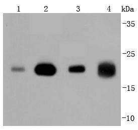
Fig1: Western blot analysis of Ras on different lysates using anti-Ras antibody at 1/1,000 dilution. Positive control: Lane 1: 293T Lane 2: MCF-7 Lane 3: Hela Lane 4: zebrafishApplication
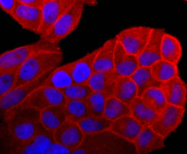
Fig2: ICC staining Ras in Hela cells (red). The nuclear counter stain is DAPI (blue). Cells were fixed in paraformaldehyde, permeabilised with 0.25% Triton X100/PBS.Application
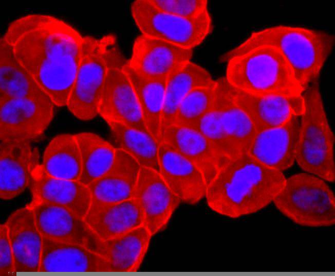
Fig3: ICC staining Ras in MCF-7 cells (red). The nuclear counter stain is DAPI (blue). Cells were fixed in paraformaldehyde, permeabilised with 0.25% Triton X100/PBS.Application
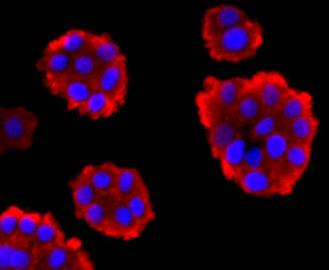
Fig4: ICC staining Ras in PC-12 cells (red). The nuclear counter stain is DAPI (blue). Cells were fixed in paraformaldehyde, permeabilised with 0.25% Triton X100/PBS.| Product Name | Ras [JF10-11] |
|---|---|
| Antibody Type | Primary Antibodies |
| Antigen Alias | C-BAS/HAS antibody c-H-ras antibody C-HA-RAS1 antibody CTLO antibody GTPase HRas antibody GTPase KRas antibody GTPase NRas antibody H-Ras-1 antibody H-RASIDX antibody Ha-Ras antibody HAMSV antibody HRAS antibody HRAS1 antibody K RAS2A antibody K RAS2B antibody K RAS4A antibody K RAS4B antibody K-RAS antibody KRAS antibody KRAS1 antibody KRAS2 antibody N-RAS antibody N-terminally processed antibody NRAS antibody NRAS1 antibody p21ras antibody RASH_HUMAN antibody RASH1 antibody RASK2 antibody Transforming protein p21 antibody v Ha ras Harvey rat sarcoma viral oncogene homolog antibody v Ki ras2 Kirsten rat sarcoma viral oncogene homolog antibody v ras neuroblastoma RAS viral oncogene homolog antibody |
| Product description | The mammalian c-H-, c-K- and N-Ras proto-oncogenes encode guanine nucleotide-binding proteins that are ubiquitously expressed in vertebrate cells. c-H- and c-K-Ras are cellular homologs of the v-H and v-K-Ras sequences originally isolated from the Harvey and Kirsten strains of rat sarcoma virus. Ras-encoded proteins bind GDP and GTP with high affinity and possess a low level intrinsic GTPase activity that can be stimulated over 100-fold by interaction with cytosolic GTPase activating protein (GAP), a potential effector for Ras p21 function. Point mutations at amino acids 12, 13, 59 and 61 within domains responsible for GTP binding and hydrolysis activate Ras proteins to their oncogenic form and block the ability of the GTPase activity to be stimulated by GAP. Several additional proteins with GAP activity have been identified and shown to interact with p21 Ras or other members of the Ras gene family. |
| Immunogen | recombinant protein |
| Clonality | Monoclonal |
|---|---|
| Isotype | IgG |
| Host Species | Recombinant rabbit |
| Tested Applications | WBICC/IFIP |
| WB:1:1,000-1:2,000 ICC:1:100-1:500 |
|
| Species Reactivity | HumanMouseRatZebra Fish |
| Concentration | 1mg/ml |
| Alternative Names | C-BAS/HAS antibody c-H-ras antibody C-HA-RAS1 antibody CTLO antibody GTPase HRas antibody GTPase KRas antibody GTPase NRas antibody H-Ras-1 antibody H-RASIDX antibody Ha-Ras antibody HAMSV antibody HRAS antibody HRAS1 antibody K RAS2A antibody K RAS2B antibody K RAS4A antibody K RAS4B antibody K-RAS antibody KRAS antibody KRAS1 antibody KRAS2 antibody N-RAS antibody N-terminally processed antibody NRAS antibody NRAS1 antibody p21ras antibody RASH_HUMAN antibody RASH1 antibody RASK2 antibody Transforming protein p21 antibody v Ha ras Harvey rat sarcoma viral oncogene homolog antibody v Ki ras2 Kirsten rat sarcoma viral oncogene homolog antibody v ras neuroblastoma RAS viral oncogene homolog antibody |
|---|---|
| Molecular Weight(MW) | 21 kDa |
| Cellular Localization | Cytoplasm, Cell membrane, Golgi apparatus, Nucleus. |
Application
Fig1: Western blot analysis of Ras on different lysates using anti-Ras antibody at 1/1,000 dilution. Positive control: Lane 1: 293T Lane 2: MCF-7 Lane 3: Hela Lane 4: zebrafish
Application
Fig2: ICC staining Ras in Hela cells (red). The nuclear counter stain is DAPI (blue). Cells were fixed in paraformaldehyde, permeabilised with 0.25% Triton X100/PBS.
Application
Fig3: ICC staining Ras in MCF-7 cells (red). The nuclear counter stain is DAPI (blue). Cells were fixed in paraformaldehyde, permeabilised with 0.25% Triton X100/PBS.
Application
Fig4: ICC staining Ras in PC-12 cells (red). The nuclear counter stain is DAPI (blue). Cells were fixed in paraformaldehyde, permeabilised with 0.25% Triton X100/PBS.| Positive Control | 293T, MCF-7, Hela, PC-12, zebrafish. |
|---|---|
| Application Notes | WB:1:1,000-1:2,000 ICC:1:100-1:500 |
| Form | Liquid |
|---|---|
| Storage Instructions | Store at +4℃ after thawing. Aliquot store at -20℃ or -80℃. Avoid repeated freeze / thaw cycles. |
| Storage Buffer | 1*TBS (pH7.4), 1%BSA, 40%Glycerol. Preservative: 0.05% Sodium Azide. |


