Application
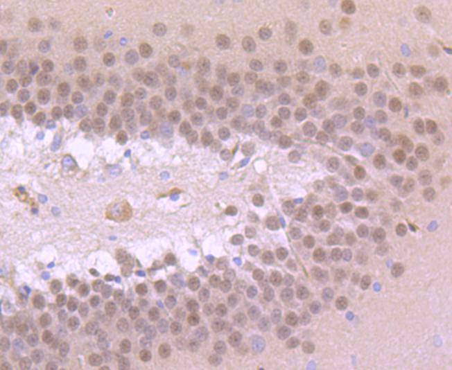
Fig1: Immunohistochemical analysis of paraffin-embedded rat brain tissue using anti-TBR1 antibody. Counter stained with hematoxylin.Application
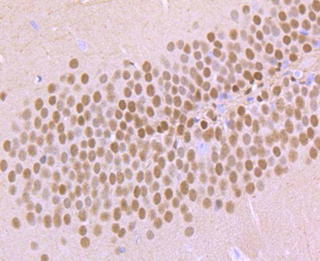
Fig2: Immunohistochemical analysis of paraffin-embedded mouse brain tissue using anti-TBR1 antibody. Counter stained with hematoxylin.Application
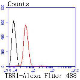
Fig3: Flow cytometric analysis of SH-SY-5Y cells with TBR1 antibody at 1/50 dilution (red) compared with an unlabelled control (cells without incubation with primary antibody; black). Alexa Fluor 488-conjugated goat anti rabbit IgG was used as the secondary antibody| Product Name | TBR1 [JF10-00] |
|---|---|
| Antibody Type | Primary Antibodies |
| Antigen Alias | T box brain 1 antibody T box brain protein 1 antibody T brain 1 protein antibody T-box brain protein 1 antibody T-brain-1 antibody TBR 1 antibody TBR-1 antibody Tbr1 antibody TBR1_HUMAN antibody TES 56 antibody TES-56 antibody |
| Product description | A novel murine and human gene, TBR-1, encodes a putative transcription factor related to the Brachyrury (T) gene that is expressed only in postmitotic cells. T-brain-1 (TBR-1) mRNA is largely restricted to the cerebral cortex, where, during embryogenesis, it defines different regions that give rise to the palecortex, limbic cortex and neocortex. TBR-1, Pax-6 and Emx-1 are expressed in the mouse and chicken pallium. The pallio-subpallial boundary lies at the interface between the TBR-1 and Dlx-2 expression domains. Chicken genes homolgous to these mouse genes are expressed in topologically comparable patterns during development, suggesting that mouse and chicken may have similar histogenetic specification processes and field homologies. CASK/LIN-2, a membrane-associated guanylate kinase, is required for EGFR localization and signaling. In adult rat brain, CASK is concentrated at neuronal synapses and binds to the cell-surface proteins. CASK can interact with TBR-1, which is involved in forebrain development. CASK enters into the nucleus and binds to a specific DNA sequence (the T-element) in a complex with TBR-1. Thus, CASK acts as a coactivator of TBR-1 to induce transcription of T-element containing genes, including reelin. |
| Immunogen | recombinant protein |
| Clonality | Monoclonal |
|---|---|
| Isotype | IgG |
| Host Species | Recombinant rabbit |
| Tested Applications | WBIHCFC |
| WB:1:500-1:1,000 FC:1:50-1:100 IHC:1:50-1:200 |
|
| Species Reactivity | HumanMouseRat |
| Concentration | 1mg/ml |
| Alternative Names | T box brain 1 antibody T box brain protein 1 antibody T brain 1 protein antibody T-box brain protein 1 antibody T-brain-1 antibody TBR 1 antibody TBR-1 antibody Tbr1 antibody TBR1_HUMAN antibody TES 56 antibody TES-56 antibody |
|---|---|
| Molecular Weight(MW) | 74 kDa |
| Cellular Localization | Nucleus. |
Application
Fig1: Immunohistochemical analysis of paraffin-embedded rat brain tissue using anti-TBR1 antibody. Counter stained with hematoxylin.
Application
Fig2: Immunohistochemical analysis of paraffin-embedded mouse brain tissue using anti-TBR1 antibody. Counter stained with hematoxylin.
Application
Fig3: Flow cytometric analysis of SH-SY-5Y cells with TBR1 antibody at 1/50 dilution (red) compared with an unlabelled control (cells without incubation with primary antibody; black). Alexa Fluor 488-conjugated goat anti rabbit IgG was used as the secondary antibody| Positive Control | Rat brain tissue, mouse brain tissue. |
|---|---|
| Application Notes | WB:1:500-1:1,000 FC:1:50-1:100 IHC:1:50-1:200 |
| Form | Liquid |
|---|---|
| Storage Instructions | Store at +4℃ after thawing. Aliquot store at -20℃ or -80℃. Avoid repeated freeze / thaw cycles. |
| Storage Buffer | 1*TBS (pH7.4), 1%BSA, 40%Glycerol. Preservative: 0.05% Sodium Azide. |


