Application
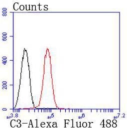
Fig3: Flow cytometric analysis of HepG2 cells with C3 antibody at 1/50 dilution (red) compared with an unlabelled control (cells without incubation with primary antibody; black). Alexa Fluor 488-conjugated goat anti rabbit IgG was used as the secondary antibodyApplication
Fig3: Flow cytometric analysis of HepG2 cells with C3 antibody at 1/50 dilution (red) compared with an unlabelled control (cells without incubation with primary antibody; black). Alexa Fluor 488-conjugated goat anti rabbit IgG was used as the secondary antibodyApplication
Fig3: Flow cytometric analysis of HepG2 cells with C3 antibody at 1/50 dilution (red) compared with an unlabelled control (cells without incubation with primary antibody; black). Alexa Fluor 488-conjugated goat anti rabbit IgG was used as the secondary antibody| Product Name | C3 [JF10-30] |
|---|---|
| Antibody Type | Primary Antibodies |
| Antigen Alias | Acylation stimulating protein cleavage product antibody AHUS5 antibody ARMD9 antibody ASP antibody C3 and PZP like alpha 2 macroglobulin domain containing protein 1 antibody C3 and PZP-like alpha-2-macroglobulin domain-containing protein 1 antibody C3 antibody CO3_HUMAN antibody Complement C3 antibody Complement C3c alpha'' chain fragment 2 antibody Complement component 3 antibody Complement factor 3 antibody CPAMD1 antibody HEL S 62p antibody |
| Product description | Complement C3 precursor contains complement C3 beta chain, complement C3 alpha chain, C3a anaphylatoxin, complement C3b alpha chain, complement C3c fragment, complement C3dg fragment, complement C3g fragment, complement C3d fragment and complement C3f fragment. C3a, C4a, and C5a are potent anaphylatoxins that are released during complement activation, a system of ligand-surface protein interactions specific to cells of hematopoietic lineage that aids in the elimination of pathogens. C3a and C5a secretion correlates with pathophysiological phenotypes such as asthma and bacterial meningitis. Binding of these proteins to their respective G protein-coupled receptors (C3aR, C5aR), which are present on the surface of myeloid leukocytes, induces proinflammatory events such as cellular degranulation, smooth muscle contraction, arachidonic acid metabolism, cytokine release, leukocyte activation and cellular chemotaxis. C3aR is expressed in brain and activated B-lymphocytes whereas C5aR is prevalent on the surface of hepatocyte, lung, smooth muscle, and endothelial cells. Upon activation, C3aR and C5aR are susceptible to rapid GRK-mediated phosphorylation and clathrin-coated vesicle targeting. C5aR utilizes the Ras-Raf-ERK1/2 cascade and couples to Gi/G16 proteins. |
| Immunogen | recombinant protein |
| Clonality | Monoclonal |
|---|---|
| Isotype | IgG |
| Host Species | Recombinant rabbit |
| Tested Applications | WBICC/IFFC |
| WB:1:1,000 FC:1:50-1:100 ICC:1:50-1:100 |
|
| Species Reactivity | Human |
| Concentration | 1mg/ml |
| Alternative Names | Acylation stimulating protein cleavage product antibody AHUS5 antibody ARMD9 antibody ASP antibody C3 and PZP like alpha 2 macroglobulin domain containing protein 1 antibody C3 and PZP-like alpha-2-macroglobulin domain-containing protein 1 antibody C3 antibody CO3_HUMAN antibody Complement C3 antibody Complement C3c alpha'' chain fragment 2 antibody Complement component 3 antibody Complement factor 3 antibody CPAMD1 antibody HEL S 62p antibody |
|---|---|
| Molecular Weight(MW) | 100 kDa |
| Cellular Localization | Secreted. |
| SwissProt ID | P01024 |
|---|
Application
Fig3: Flow cytometric analysis of HepG2 cells with C3 antibody at 1/50 dilution (red) compared with an unlabelled control (cells without incubation with primary antibody; black). Alexa Fluor 488-conjugated goat anti rabbit IgG was used as the secondary antibody
Application
Fig3: Flow cytometric analysis of HepG2 cells with C3 antibody at 1/50 dilution (red) compared with an unlabelled control (cells without incubation with primary antibody; black). Alexa Fluor 488-conjugated goat anti rabbit IgG was used as the secondary antibody
Application
Fig3: Flow cytometric analysis of HepG2 cells with C3 antibody at 1/50 dilution (red) compared with an unlabelled control (cells without incubation with primary antibody; black). Alexa Fluor 488-conjugated goat anti rabbit IgG was used as the secondary antibody| Positive Control | NIH/3T3, Hela, HepG2. |
|---|---|
| Application Notes | WB:1:1,000 FC:1:50-1:100 ICC:1:50-1:100 |
| Form | Liquid |
|---|---|
| Storage Instructions | Store at +4℃ after thawing. Aliquot store at -20℃ or -80℃. Avoid repeated freeze / thaw cycles. |
| Storage Buffer | 1*TBS (pH7.4), 1%BSA, 40%Glycerol. Preservative: 0.05% Sodium Azide. |


