WB
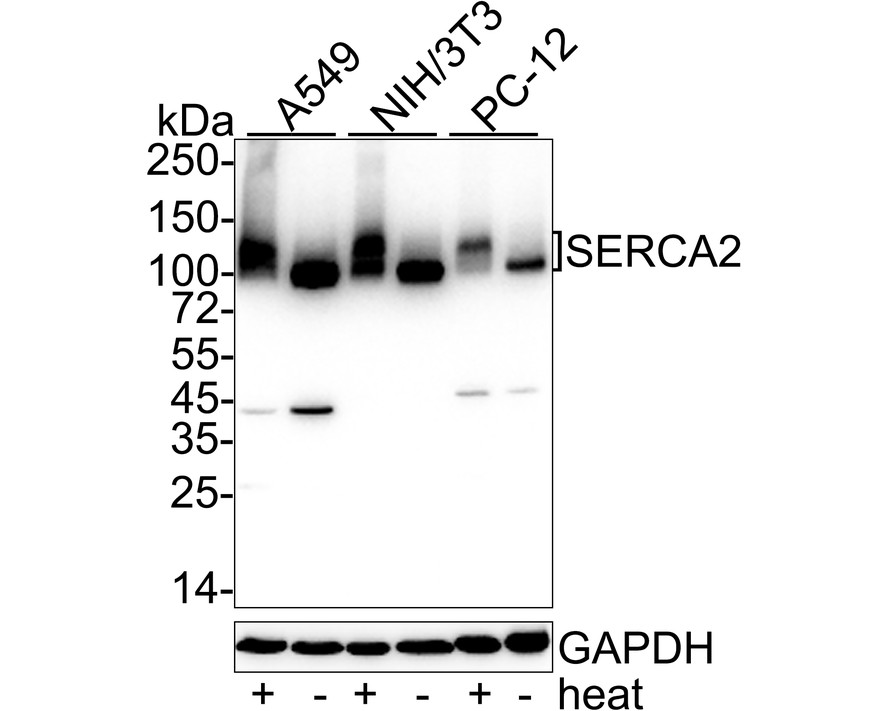
Western blot analysis of SERCA2 on different lysates with Rabbit anti-SERCA2 antibody at 1/1,000 dilution. Lane 1: A549 cell lysate, Lane 2: A549 cell lysate (no heat), Lane 3: NIH/3T3 cell lysate, Lane 4: NIH/3T3 cell lysate (no heat), Lane 5: PC-12 cell lysate, Lane 6: PC-12 cell lysate (no heat), Notice: no heat means the lysate is not boiled. Lysates/proteins at 20 µg/Lane. Exposure time: 25 seconds; 4-20% SDS-PAGE gel. Proteins were transferred to a PVDF membrane and blocked with 5% NFDM/TBST for 1 hour at room temperature. The primary antibody at 1/1,000 dilution was used in 5% NFDM/TBST at 4℃ overnight. Goat Anti-Rabbit IgG - HRP Secondary Antibody at 1/50,000 dilution was used for 1 hour at room temperature.ICC/IF
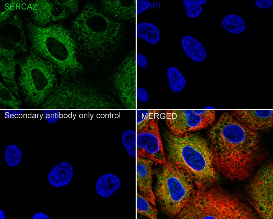
Immunocytochemistry analysis of A549 cells labeling SERCA2 with Rabbit anti-SERCA2 antibody at 1/100 dilution. Cells were fixed in 4% paraformaldehyde for 15 minutes at room temperature, permeabilized with 0.1% Triton X-100 in PBS for 15 minutes at room temperature, then blocked with 1% BSA in 10% negative goat serum for 1 hour at room temperature. Cells were then incubated with Rabbit anti-SERCA2 antibody at 1/100 dilution in 1% BSA in PBST overnight at 4 ℃. Goat Anti-Rabbit IgG H&L (488) was used as the secondary antibody at 1/1,000 dilution. PBS instead of the primary antibody was used as the secondary antibody only control. Nuclear DNA was labelled in blue with DAPI. Beta tubulin (red) was stained at 1/100 dilution overnight at +4℃. Goat Anti-Mouse IgG H&L (594) was used as the secondary antibody at 1/1,000 dilution.ICC/IF
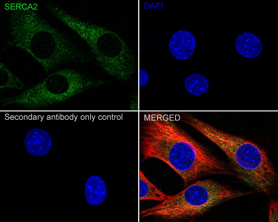
Immunocytochemistry analysis of NIH/3T3 cells labeling SERCA2 with Rabbit anti-SERCA2 antibody at 1/100 dilution. Cells were fixed in 4% paraformaldehyde for 15 minutes at room temperature, permeabilized with 0.1% Triton X-100 in PBS for 15 minutes at room temperature, then blocked with 1% BSA in 10% negative goat serum for 1 hour at room temperature. Cells were then incubated with Rabbit anti-SERCA2 antibody at 1/100 dilution in 1% BSA in PBST overnight at 4 ℃. Goat Anti-Rabbit IgG H&L (488) was used as the secondary antibody at 1/1,000 dilution. PBS instead of the primary antibody was used as the secondary antibody only control. Nuclear DNA was labelled in blue with DAPI. Beta tubulin (red) was stained at 1/100 dilution overnight at +4℃. Goat Anti-Mouse IgG H&L (594) was used as the secondary antibody at 1/1,000 dilution.IHC
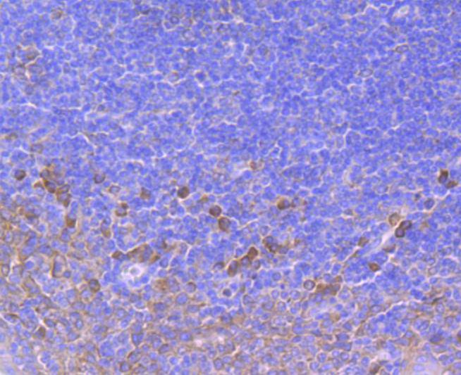
Immunohistochemical analysis of paraffin-embedded human tonsil tissue using anti-SERCA2 antibody. Counter stained with hematoxylin.IHC
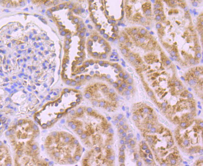
Immunohistochemical analysis of paraffin-embedded human kidney tissue using anti-SERCA2 antibody. Counter stained with hematoxylin.IHC
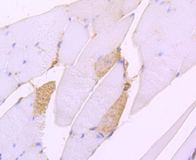
Immunohistochemical analysis of paraffin-embedded mouse skeletal muscle tissue using anti-SERCA2 antibody. Counter stained with hematoxylin.IHC
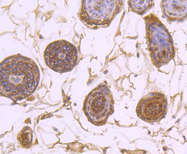
Immunohistochemical analysis of paraffin-embedded mouse skin tissue using anti-SERCA2 antibody. Counter stained with hematoxylin.IHC
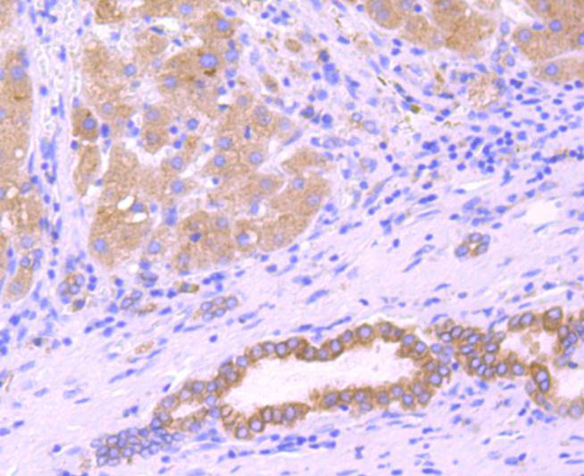
Immunohistochemical analysis of paraffin-embedded human liver cancer tissue using anti-SERCA2 antibody. Counter stained with hematoxylin.IHC
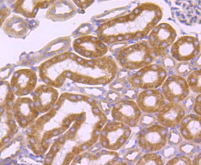
Immunohistochemical analysis of paraffin-embedded mouse kidney tissue using anti-SERCA2 antibody. Counter stained with hematoxylin.IHC
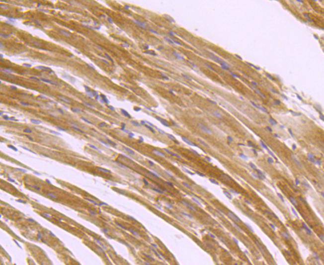
Immunohistochemical analysis of paraffin-embedded mouse heart tissue using anti-SERCA2 antibody. Counter stained with hematoxylin.FC
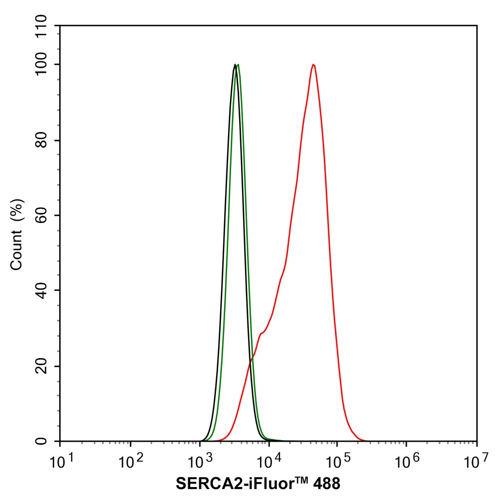
Flow cytometric analysis of A549 cells labeling SERCA2. Cells were fixed and permeabilized. Then stained with the primary antibody (1/1,000) (red) compared with Rabbit IgG Isotype Control (green). After incubation of the primary antibody at +4℃ for an hour, the cells were stained with a 488 conjugate-Goat anti-Rabbit IgG Secondary antibody at 1/1,000 dilution for 30 minutes at +4℃. Unlabelled sample was used as a control (cells without incubation with primary antibody; black).| Product Name | SERCA2 Recombinant Rabbit Monoclonal Antibody |
|---|---|
| Antibody Type | Primary Antibodies |
| Product description | ATP dependent calcium pumps are responsible, in part, for the maintenance of low cytoplasmic free calcium concentrations. The ATP pumps that reside in intracellular organelles are encoded by a family of structurally related enzymes, termed the sarcoplasmic or endoplasmic reticulum calcium (SERCA) ATPases. The sarcoplasmic reticulum of striated muscle is a specialized intracellular membrane system that plays a critical role in the contraction and relaxation of muscle. The SERCAs mediate Ca2+ uptake into intracellular stores. SERCA-mediated Ca2+ uptake induces and maintains muscular relaxation. The SERCA1 gene is exclusively expressed in type II (fast) skeletal muscle. The SERCA2 gene is subject to tissue-dependent processing which is responsible for the generation of the SERCA2a muscle-specific form expressed in type I (slow) skeletal, cardiac and smooth muscle, and the SERCA2b isoform expressed in all cell types. The SERCA3 gene is not as well characterized and is found in non-muscle cells. SERCA2 plays an important part in regulating cardiac contractile function. SERCA3 is an isoform expressed in several cell types including platelets, lymphoid cells and mast cells. SERCA1, SERCA2 and SERCA3 all undergo alternative splicing. |
| Immunogen | Synthetic peptide within Human SERCA2 aa 999-1,042 / 1,042. |
| Clonality | monoclonal |
|---|---|
| Isotype | IgG |
| Host Species | Recombinant rabbit |
| Tested Applications | FCICC/IFIHCWB |
| WB:1:1000 IHC:1:50-1:500 ICC/IF:1:100 FC:1:1000 |
|
| Species Reactivity | HumanMouseRat |
| Concentration | 1mg/ml |
| Purification | Protein A |
| Alternative Names | AT2A2_HUMAN antibody Atp2a2 antibody ATP2B antibody ATPase Ca++ transporting cardiac muscle slow twitch 2 antibody Calcium pump 2 antibody Calcium-transporting ATPase sarcoplasmic reticulum type antibody Calcium-transporting ATPase sarcoplasmic reticulum type slow twitch skeletal muscle isoform antibody Cardiac Ca2+ ATPase antibody DAR antibody DD antibody Endoplasmic reticulum class 1/2 Ca(2+) ATPase antibody MGC45367 antibody Sarcoplasmic/endoplasmic reticulum calcium ATPase 2 antibody SERCA 2 antibody SERCA2 antibody serca2a antibody slow twitch skeletal muscle isoform antibody SR Ca(2+)-ATPase 2 antibody |
|---|---|
| Molecular Weight(MW) | 115 kDa |
| Cellular Localization | Endoplasmic reticulum membrane, Sarcoplasmic reticulum membrane. |
WB
Western blot analysis of SERCA2 on different lysates with Rabbit anti-SERCA2 antibody at 1/1,000 dilution. Lane 1: A549 cell lysate, Lane 2: A549 cell lysate (no heat), Lane 3: NIH/3T3 cell lysate, Lane 4: NIH/3T3 cell lysate (no heat), Lane 5: PC-12 cell lysate, Lane 6: PC-12 cell lysate (no heat), Notice: no heat means the lysate is not boiled. Lysates/proteins at 20 µg/Lane. Exposure time: 25 seconds; 4-20% SDS-PAGE gel. Proteins were transferred to a PVDF membrane and blocked with 5% NFDM/TBST for 1 hour at room temperature. The primary antibody at 1/1,000 dilution was used in 5% NFDM/TBST at 4℃ overnight. Goat Anti-Rabbit IgG - HRP Secondary Antibody at 1/50,000 dilution was used for 1 hour at room temperature.
ICC/IF
Immunocytochemistry analysis of A549 cells labeling SERCA2 with Rabbit anti-SERCA2 antibody at 1/100 dilution. Cells were fixed in 4% paraformaldehyde for 15 minutes at room temperature, permeabilized with 0.1% Triton X-100 in PBS for 15 minutes at room temperature, then blocked with 1% BSA in 10% negative goat serum for 1 hour at room temperature. Cells were then incubated with Rabbit anti-SERCA2 antibody at 1/100 dilution in 1% BSA in PBST overnight at 4 ℃. Goat Anti-Rabbit IgG H&L (488) was used as the secondary antibody at 1/1,000 dilution. PBS instead of the primary antibody was used as the secondary antibody only control. Nuclear DNA was labelled in blue with DAPI. Beta tubulin (red) was stained at 1/100 dilution overnight at +4℃. Goat Anti-Mouse IgG H&L (594) was used as the secondary antibody at 1/1,000 dilution.
ICC/IF
Immunocytochemistry analysis of NIH/3T3 cells labeling SERCA2 with Rabbit anti-SERCA2 antibody at 1/100 dilution. Cells were fixed in 4% paraformaldehyde for 15 minutes at room temperature, permeabilized with 0.1% Triton X-100 in PBS for 15 minutes at room temperature, then blocked with 1% BSA in 10% negative goat serum for 1 hour at room temperature. Cells were then incubated with Rabbit anti-SERCA2 antibody at 1/100 dilution in 1% BSA in PBST overnight at 4 ℃. Goat Anti-Rabbit IgG H&L (488) was used as the secondary antibody at 1/1,000 dilution. PBS instead of the primary antibody was used as the secondary antibody only control. Nuclear DNA was labelled in blue with DAPI. Beta tubulin (red) was stained at 1/100 dilution overnight at +4℃. Goat Anti-Mouse IgG H&L (594) was used as the secondary antibody at 1/1,000 dilution.
IHC
Immunohistochemical analysis of paraffin-embedded human tonsil tissue using anti-SERCA2 antibody. Counter stained with hematoxylin.
IHC
Immunohistochemical analysis of paraffin-embedded human kidney tissue using anti-SERCA2 antibody. Counter stained with hematoxylin.
IHC
Immunohistochemical analysis of paraffin-embedded mouse skeletal muscle tissue using anti-SERCA2 antibody. Counter stained with hematoxylin.
IHC
Immunohistochemical analysis of paraffin-embedded mouse skin tissue using anti-SERCA2 antibody. Counter stained with hematoxylin.
IHC
Immunohistochemical analysis of paraffin-embedded human liver cancer tissue using anti-SERCA2 antibody. Counter stained with hematoxylin.
IHC
Immunohistochemical analysis of paraffin-embedded mouse kidney tissue using anti-SERCA2 antibody. Counter stained with hematoxylin.
IHC
Immunohistochemical analysis of paraffin-embedded mouse heart tissue using anti-SERCA2 antibody. Counter stained with hematoxylin.
FC
Flow cytometric analysis of A549 cells labeling SERCA2. Cells were fixed and permeabilized. Then stained with the primary antibody (1/1,000) (red) compared with Rabbit IgG Isotype Control (green). After incubation of the primary antibody at +4℃ for an hour, the cells were stained with a 488 conjugate-Goat anti-Rabbit IgG Secondary antibody at 1/1,000 dilution for 30 minutes at +4℃. Unlabelled sample was used as a control (cells without incubation with primary antibody; black).| Application Notes | WB:1:1000 IHC:1:50-1:500 ICC/IF:1:100 FC:1:1000 |
|---|
| Form | Liquid |
|---|---|
| Storage Instructions | Store at +4℃ after thawing. Aliquot store at -20℃ or -80℃. Avoid repeated freeze / thaw cycles. |
| Storage Buffer | 1*TBS (pH7.4), 1%BSA, 40%Glycerol. Preservative: 0.05% Sodium Azide. |


