Application
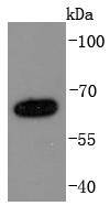
Fig1: Western blot analysis of NUMB on A431 cells lysates using anti-NUMB antibody at 1/1,000 dilution.Application
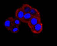
Fig2: ICC staining NUMB in Hela cells (red). The nuclear counter stain is DAPI (blue). Cells were fixed in paraformaldehyde, permeabilised with 0.25% Triton X100/PBS.Application
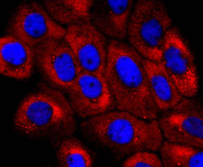
Fig3: ICC staining NUMB in A431 cells (red). The nuclear counter stain is DAPI (blue). Cells were fixed in paraformaldehyde, permeabilised with 0.25% Triton X100/PBS.Application
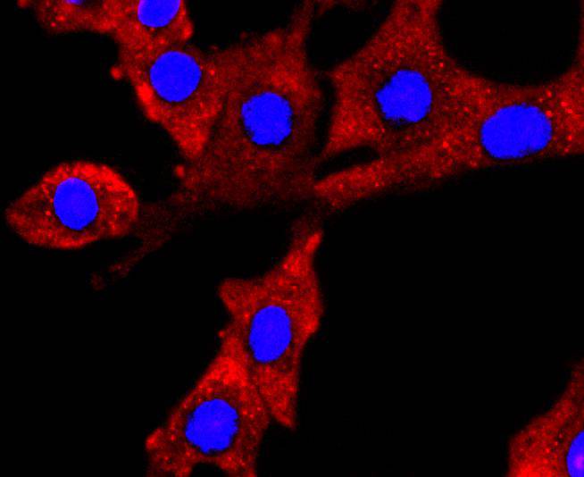
Fig4: ICC staining NUMB in NIH/3T3 cells (red). The nuclear counter stain is DAPI (blue). Cells were fixed in paraformaldehyde, permeabilised with 0.25% Triton X100/PBS.Application
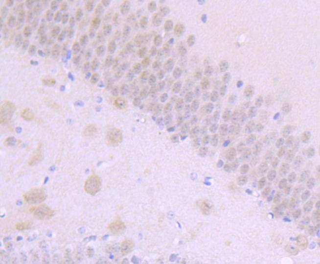
Fig5: Immunohistochemical analysis of paraffin-embedded mouse brain tissue using anti-NUMB antibody. Counter stained with hematoxylin.Application
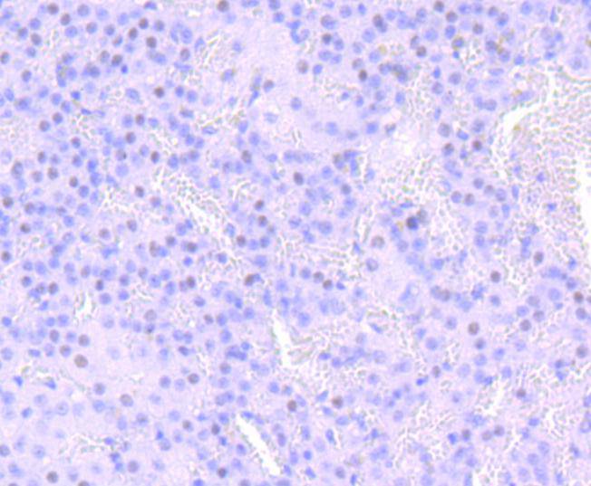
Fig6: Immunohistochemical analysis of paraffin-embedded rat adrenal gland tissue using anti-NUMB antibody. Counter stained with hematoxylin.Application
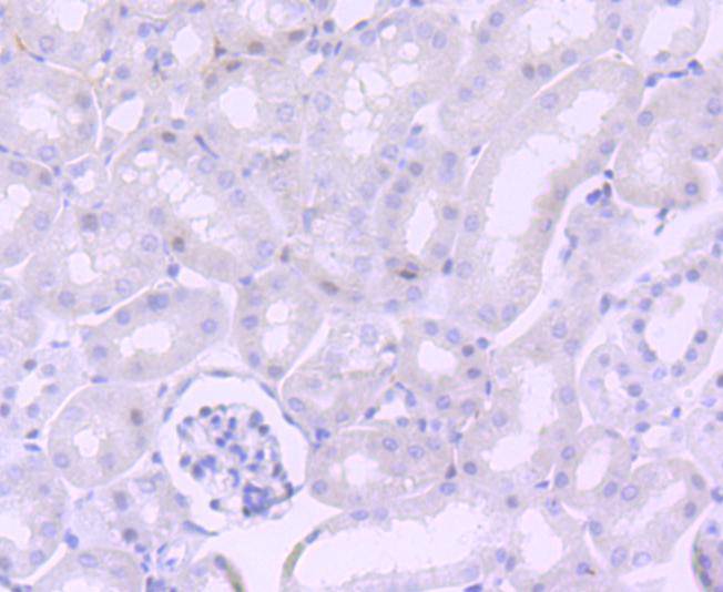
Fig7: Immunohistochemical analysis of paraffin-embedded mouse kidney tissue using anti-NUMB antibody. Counter stained with hematoxylin.| Product Name | NUMB [JM10-023] |
|---|---|
| Antibody Type | Primary Antibodies |
| Antigen Alias | c14_5527 antibody C14orf41 antibody FLJ31314 antibody h Numb antibody h-Numb antibody Numb antibody Numb homolog (Drosophila) antibody Numb homolog antibody Numb protein homolog antibody NUMB_HUMAN antibody Protein numb homolog antibody Protein S171 antibody S171 antibody |
| Product description | Neuronal cell fate decisions are directed in Drosophila by NUMB, a signaling adapter protein with two protein-protein interaction domains: a phosphotyrosine-binding domain and a proline-rich SH3-binding region (PRR). Mammalian NUMB homologs play a role in the determination of cell fates during development and bind with EPS15, LNX1, and NOTCH1. Conditional mouse mutants with deletion of NUMB in developing sensory ganglia show a reduction in axonal arborization in afferent fibers. Changes in cellular calcium homeostasis influences NUMB-dependent cell fate decisions during development of the nervous system. Chicken NUMB (c-NUMB) protein is localized to the basal cortex of mitotic neuroepithelial cells. |
| Immunogen | recombinant protein |
| Clonality | Monoclonal |
|---|---|
| Isotype | IgG |
| Host Species | Recombinant rabbit |
| Tested Applications | WBICCIHC |
| WB:1:1,000-1:5,000 ICC:1:50-1:200 IHC:1:50-1:100 |
|
| Species Reactivity | HumanMouseRat |
| Concentration | 1mg/ml |
| Alternative Names | c14_5527 antibody C14orf41 antibody FLJ31314 antibody h Numb antibody h-Numb antibody Numb antibody Numb homolog (Drosophila) antibody Numb homolog antibody Numb protein homolog antibody NUMB_HUMAN antibody Protein numb homolog antibody Protein S171 antibody S171 antibody |
|---|---|
| Molecular Weight(MW) | 71 kDa |
| Cellular Localization | Membrane. |
Application
Fig1: Western blot analysis of NUMB on A431 cells lysates using anti-NUMB antibody at 1/1,000 dilution.
Application
Fig2: ICC staining NUMB in Hela cells (red). The nuclear counter stain is DAPI (blue). Cells were fixed in paraformaldehyde, permeabilised with 0.25% Triton X100/PBS.
Application
Fig3: ICC staining NUMB in A431 cells (red). The nuclear counter stain is DAPI (blue). Cells were fixed in paraformaldehyde, permeabilised with 0.25% Triton X100/PBS.
Application
Fig4: ICC staining NUMB in NIH/3T3 cells (red). The nuclear counter stain is DAPI (blue). Cells were fixed in paraformaldehyde, permeabilised with 0.25% Triton X100/PBS.
Application
Fig5: Immunohistochemical analysis of paraffin-embedded mouse brain tissue using anti-NUMB antibody. Counter stained with hematoxylin.
Application
Fig6: Immunohistochemical analysis of paraffin-embedded rat adrenal gland tissue using anti-NUMB antibody. Counter stained with hematoxylin.
Application
Fig7: Immunohistochemical analysis of paraffin-embedded mouse kidney tissue using anti-NUMB antibody. Counter stained with hematoxylin.| Positive Control | A431, NIH/3T3, Hela, rat adrenal gland tissue, mouse kidney tissue, mouse brain tissue. |
|---|---|
| Application Notes | WB:1:1,000-1:5,000 ICC:1:50-1:200 IHC:1:50-1:100 |
| Form | Liquid |
|---|---|
| Storage Instructions | Store at +4℃ after thawing. Aliquot store at -20℃ or -80℃. Avoid repeated freeze / thaw cycles. |
| Storage Buffer | 1*TBS (pH7.4), 1%BSA, 40%Glycerol. Preservative: 0.05% Sodium Azide. |


