WB
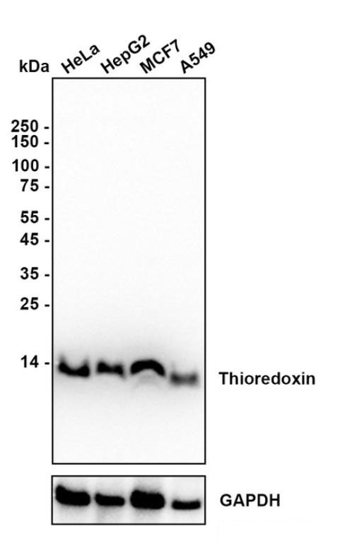
Western blot analysis of Thioredoxin on different lysates with Rabbit anti-Thioredoxin antibody at 1/5,000 dilution. Lane 1: HeLa (Human cervical adenocarcinoma cells) cell lysate, Lane 2: Hep G2 (Human liver cancer cells) cell lysate, Lane 3: MCF7 (Human breast cancer cells) cell lysate, Lane 4: A549 (Human lung adenocarcinoma cells) cell lysate, Lysates/proteins at 15 µg/Lane. Exposure time: 18 seconds; Blocking: 5% NFDM/TBST, 1 hour at room temperature Primary antibody: 1/5,000 in primary antibody dilution buffer, overnight at 4 ℃ Secondary antibody: Goat anti-Rabbit IgG-HRP, 1/50,000 in 5% NFDM/TBST, 1 hour at room temperature.ICC/IF
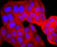
ICC staining TRX1 in Hela cells (red). The nuclear counter stain is DAPI (blue). Cells were fixed in paraformaldehyde, permeabilised with 0.25% Triton X100/PBS.ICC/IF
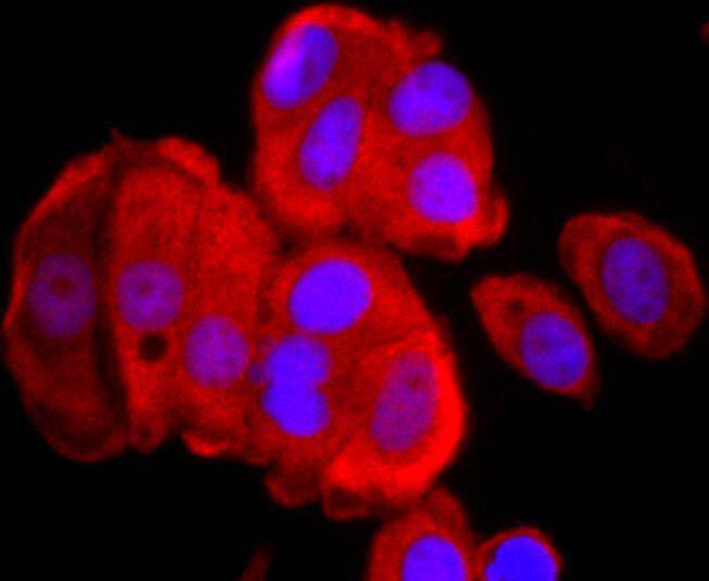
Fig3: ICC staining TRX1 in MCF-7 cells (red). The nuclear counter stain is DAPI (blue). Cells were fixed in paraformaldehyde, permeabilised with 0.25% Triton X100/PBS.ICC/IF
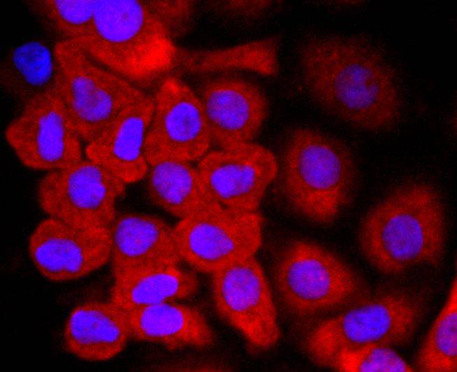
ICC staining TRX1 in SKOV-3 cells (red). The nuclear counter stain is DAPI (blue). Cells were fixed in paraformaldehyde, permeabilised with 0.25% Triton X100/PBS.IHC
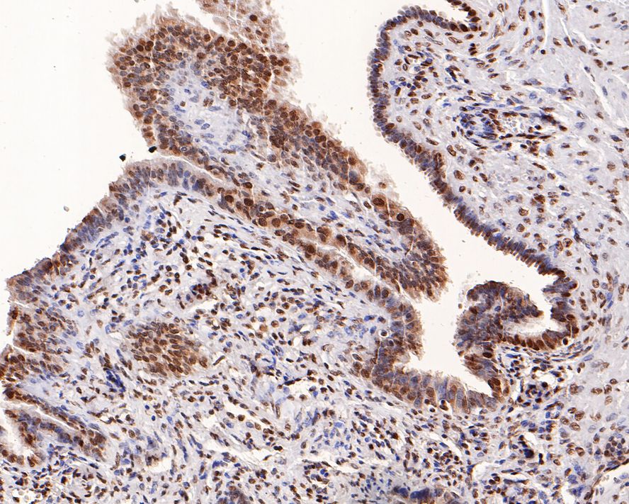
Immunohistochemical analysis of paraffin-embedded human fallopian tube tissue with Rabbit anti-Thioredoxin antibody at 1/5,000 dilution. The section was pre-treated using heat mediated antigen retrieval with sodium citrate buffer (pH 6.0) (high pressure) for 2 minutes. The tissues were blocked in 1% BSA for 20 minutes at room temperature, washed with ddH2O and PBS, and then probed with the primary antibody at 1/5,000 dilution for 1 hour at room temperature. The detection was performed using an HRP conjugated compact polymer system. DAB was used as the chromogen. Tissues were counterstained with hematoxylin and mounted with DPX.IHC
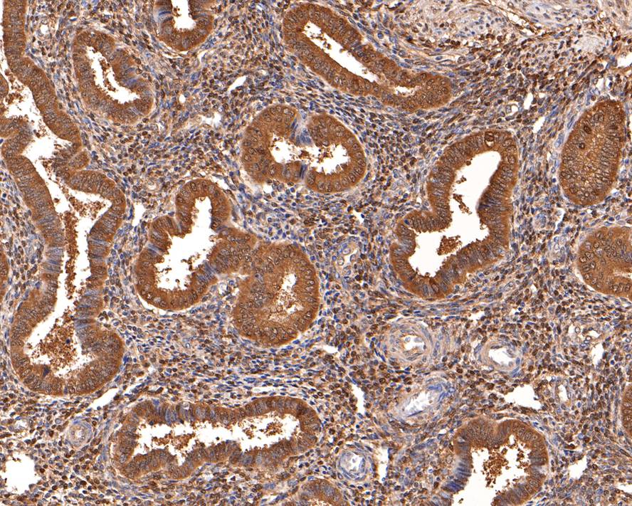
Immunohistochemical analysis of paraffin-embedded human uterus tissue with Rabbit anti-Thioredoxin antibody at 1/400 dilution. The section was pre-treated using heat mediated antigen retrieval with sodium citrate buffer (pH 6.0) (high pressure) for 2 minutes. The tissues were blocked in 1% BSA for 20 minutes at room temperature, washed with ddH2O and PBS, and then probed with the primary antibody at 1/400 dilution for 1 hour at room temperature. The detection was performed using an HRP conjugated compact polymer system. DAB was used as the chromogen. Tissues were counterstained with hematoxylin and mounted with DPX.IHC
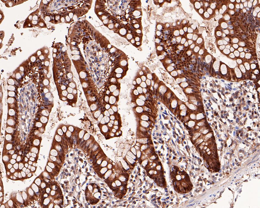
Immunohistochemical analysis of paraffin-embedded human small intestine tissue with Rabbit anti-Thioredoxin antibody at 1/2,000 dilution. The section was pre-treated using heat mediated antigen retrieval with sodium citrate buffer (pH 6.0) (high pressure) for 2 minutes. The tissues were blocked in 1% BSA for 20 minutes at room temperature, washed with ddH2O and PBS, and then probed with the primary antibody at 1/2,000 dilution for 1 hour at room temperature. The detection was performed using an HRP conjugated compact polymer system. DAB was used as the chromogen. Tissues were counterstained with hematoxylin and mounted with DPX.| Product Name | Thioredoxin Recombinant Rabbit Monoclonal Antibody |
|---|---|
| Antibody Type | Primary Antibodies |
| Product description | Thioredoxin (Trx) is a redox protein that is found in several species, such as bacteria, plants and mammals, and contains a conserved active site, consisting of Trp-Cys-Gly-Pro-Cys. Trx has several biological functions. It acts as a hydrogen donor for ribonucleotide reductase, which is critical for DNA synthesis, and modulates the DNA-binding activity of several transcription factors, including NFkB, AP-1, p53, TFIIIC and glucocorticoid receptor. Trx also stimulates cell growth, is an inhibitor of apoptosis and plays a role in the protection against oxidative stress. Drugs that inhibit Trx have antitumor activity, suggesting that Trx is involved in a variety of human diseases, including cancer. TrxR is a ubiquitously expressed flavoprotein that catalyzes the NADPH-dependent reduction of Trx as well as several other oxidized cellular components. |
| Immunogen | Synthetic peptide within Human Thioredoxin aa 6-50 / 105. |
| Clonality | Monoclonal |
|---|---|
| Isotype | IgG |
| Host Species | Recombinant rabbit |
| Tested Applications | ICC/IFIHCWB |
| WB:1:5000 ICC/IF:1:100-1:200 IHC:1:400 |
|
| Species Reactivity | HumanRat |
| Concentration | 1mg/ml |
| Purification | Protein A |
| Alternative Names | ADF antibody ATL derived factor antibody ATL-derived factor antibody DKFZp686B1993 antibody MGC61975 antibody SASP antibody Surface associated sulphydryl protein antibody Surface-associated sulphydryl protein antibody testicular tissue protein Li 199 antibody THIO_HUMAN antibody Thioredoxin antibody thioredoxin delta 3 antibody TRDX antibody TRX 1 antibody Trx antibody TRX1 antibody TXN antibody TXN delta 3 antibody TXN protein antibody zgc:92903 antibody |
|---|---|
| Molecular Weight(MW) | 12 kDa |
| Cellular Localization | Nucleus, Cytoplasm, Secreted. |
| SwissProt ID | P10599 |
|---|
WB
Western blot analysis of Thioredoxin on different lysates with Rabbit anti-Thioredoxin antibody at 1/5,000 dilution. Lane 1: HeLa (Human cervical adenocarcinoma cells) cell lysate, Lane 2: Hep G2 (Human liver cancer cells) cell lysate, Lane 3: MCF7 (Human breast cancer cells) cell lysate, Lane 4: A549 (Human lung adenocarcinoma cells) cell lysate, Lysates/proteins at 15 µg/Lane. Exposure time: 18 seconds; Blocking: 5% NFDM/TBST, 1 hour at room temperature Primary antibody: 1/5,000 in primary antibody dilution buffer, overnight at 4 ℃ Secondary antibody: Goat anti-Rabbit IgG-HRP, 1/50,000 in 5% NFDM/TBST, 1 hour at room temperature.
ICC/IF
ICC staining TRX1 in Hela cells (red). The nuclear counter stain is DAPI (blue). Cells were fixed in paraformaldehyde, permeabilised with 0.25% Triton X100/PBS.
ICC/IF
Fig3: ICC staining TRX1 in MCF-7 cells (red). The nuclear counter stain is DAPI (blue). Cells were fixed in paraformaldehyde, permeabilised with 0.25% Triton X100/PBS.
ICC/IF
ICC staining TRX1 in SKOV-3 cells (red). The nuclear counter stain is DAPI (blue). Cells were fixed in paraformaldehyde, permeabilised with 0.25% Triton X100/PBS.
IHC
Immunohistochemical analysis of paraffin-embedded human fallopian tube tissue with Rabbit anti-Thioredoxin antibody at 1/5,000 dilution. The section was pre-treated using heat mediated antigen retrieval with sodium citrate buffer (pH 6.0) (high pressure) for 2 minutes. The tissues were blocked in 1% BSA for 20 minutes at room temperature, washed with ddH2O and PBS, and then probed with the primary antibody at 1/5,000 dilution for 1 hour at room temperature. The detection was performed using an HRP conjugated compact polymer system. DAB was used as the chromogen. Tissues were counterstained with hematoxylin and mounted with DPX.
IHC
Immunohistochemical analysis of paraffin-embedded human uterus tissue with Rabbit anti-Thioredoxin antibody at 1/400 dilution. The section was pre-treated using heat mediated antigen retrieval with sodium citrate buffer (pH 6.0) (high pressure) for 2 minutes. The tissues were blocked in 1% BSA for 20 minutes at room temperature, washed with ddH2O and PBS, and then probed with the primary antibody at 1/400 dilution for 1 hour at room temperature. The detection was performed using an HRP conjugated compact polymer system. DAB was used as the chromogen. Tissues were counterstained with hematoxylin and mounted with DPX.
IHC
Immunohistochemical analysis of paraffin-embedded human small intestine tissue with Rabbit anti-Thioredoxin antibody at 1/2,000 dilution. The section was pre-treated using heat mediated antigen retrieval with sodium citrate buffer (pH 6.0) (high pressure) for 2 minutes. The tissues were blocked in 1% BSA for 20 minutes at room temperature, washed with ddH2O and PBS, and then probed with the primary antibody at 1/2,000 dilution for 1 hour at room temperature. The detection was performed using an HRP conjugated compact polymer system. DAB was used as the chromogen. Tissues were counterstained with hematoxylin and mounted with DPX.| Application Notes | WB:1:5000 ICC/IF:1:100-1:200 IHC:1:400 |
|---|
| Form | Liquid |
|---|---|
| Storage Instructions | Store at +4℃ after thawing. Aliquot store at -20℃ or -80℃. Avoid repeated freeze / thaw cycles. |
| Storage Buffer | 1*TBS (pH7.4), 1%BSA, 40%Glycerol. Preservative: 0.05% Sodium Azide. |


