Application
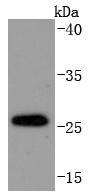
Fig1: Western blot analysis of DDIT3 on PC-12 cells lysates using anti-DDIT3 antibody at 1/1,000 dilution.Application
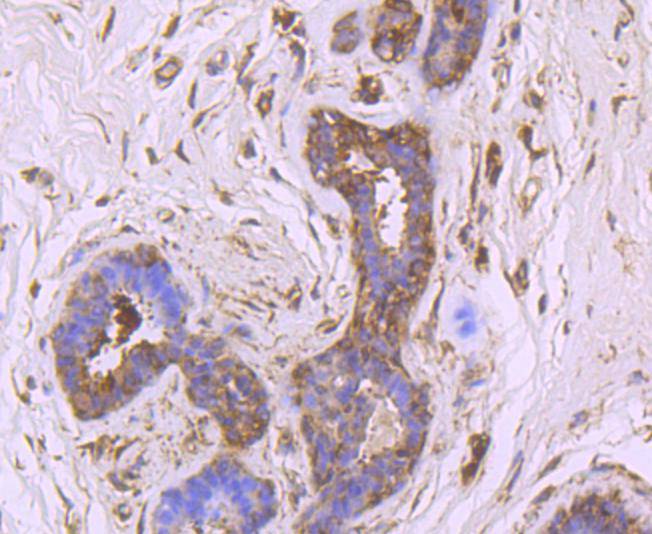
Fig2: Immunohistochemical analysis of paraffin-embedded human breast carcinoma tissue using anti-DDIT3 antibody. Counter stained with hematoxylin.Application
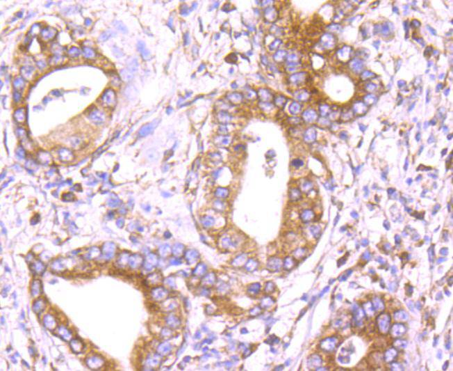
Fig3: Immunohistochemical analysis of paraffin-embedded human gastric carcinoma tissue using anti-DDIT3 antibody. Counter stained with hematoxylin.Application
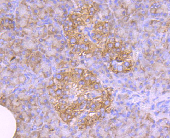
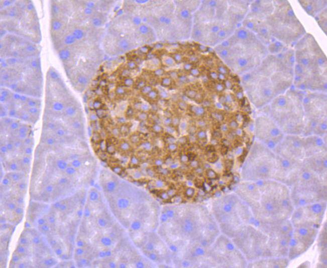
Fig4: Immunohistochemical analysis of paraffin-embedded human pancreas tissue using anti-DDIT3 antibody. Counter stained with hematoxylin.Application
Fig5: Immunohistochemical analysis of paraffin-embedded mouse pancreas tissue using anti-DDIT3 antibody. Counter stained with hematoxylin.Application
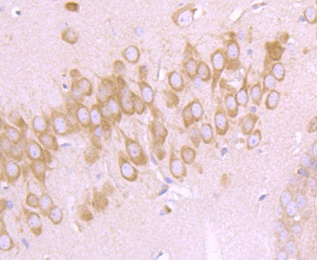
Fig6: Immunohistochemical analysis of paraffin-embedded mouse brain tissue using anti-DDIT3 antibody. Counter stained with hematoxylin.Application
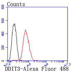
Fig7: Flow cytometric analysis of Hela cells with DDIT3 antibody at 1/50 dilution (red) compared with an unlabelled control (cells without incubation with primary antibody; black). Alexa Fluor 488-conjugated goat anti rabbit IgG was used as the secondary antibody.| Product Name | DDIT3 [JM10-31] |
|---|---|
| Antibody Type | Primary Antibodies |
| Antigen Alias | C/EBP homologous protein antibody C/EBP Homology Protein antibody C/EBP zeta antibody C/EBP-homologous protein 10 antibody C/EBP-homologous protein antibody CCAAT/enhancer binding protein homologous protein antibody CEBPZ antibody CHOP 10 antibody CHOP antibody CHOP-10 antibody CHOP10 antibody DDIT 3 antibody DDIT-3 antibody Ddit3 antibody DDIT3_HUMAN antibody DNA Damage Inducible Transcript 3 antibody DNA damage-inducible transcript 3 protein antibody GADD 153 antibody GADD153 antibody Growth Arrest and DNA Damage Inducible Protein 153 antibody Growth arrest and DNA damage inducible protein GADD153 antibody Growth arrest and DNA damage-inducible protein GADD153 antibody MGC4154 antibody |
| Product description | GADD 153 has been described as a growth arrest and DNA damage-inducible gene that encodes a C/EBP-related nuclear protein. This protein has also been designated C/EBP-homologous protein (CHOP-10). GADD 153 expression is induced by a variety of cellular stresses, inducing nutrient deprivation and metabolic perturbations. GADD 153 functions to block cells in G1 to S phase in cell cycle progression and acts by dimerizing with other C/EBP proteins to direct GADD 153 dimers away from "classical" C/EBP binding sites, recognizing instead unique "nonclassical" sites. Thus GADD 153 acts as a negative modulator of C/EBP-like proteins in certain terminally differentiated cells, similar to the regulatory function of Id on the activity of Myo D and Myo D-related proteins involved in the development of muscle cells. |
| Immunogen | recombinant protein |
| Clonality | Monoclonal |
|---|---|
| Isotype | IgG |
| Host Species | Recombinant rabbit |
| Tested Applications | WBIHCFC |
| WB:1:1,000 FC:1:50-1:100 IHC:1:50-1:200 |
|
| Species Reactivity | HumanMouseRat |
| Concentration | 1mg/ml |
| Alternative Names | C/EBP homologous protein antibody C/EBP Homology Protein antibody C/EBP zeta antibody C/EBP-homologous protein 10 antibody C/EBP-homologous protein antibody CCAAT/enhancer binding protein homologous protein antibody CEBPZ antibody CHOP 10 antibody CHOP antibody CHOP-10 antibody CHOP10 antibody DDIT 3 antibody DDIT-3 antibody Ddit3 antibody DDIT3_HUMAN antibody DNA Damage Inducible Transcript 3 antibody DNA damage-inducible transcript 3 protein antibody GADD 153 antibody GADD153 antibody Growth Arrest and DNA Damage Inducible Protein 153 antibody Growth arrest and DNA damage inducible protein GADD153 antibody Growth arrest and DNA damage-inducible protein GADD153 antibody MGC4154 antibody |
|---|---|
| Molecular Weight(MW) | 25 kDa |
| Cellular Localization | Cytoplasm, Nucleus. |
Application
Fig1: Western blot analysis of DDIT3 on PC-12 cells lysates using anti-DDIT3 antibody at 1/1,000 dilution.
Application
Fig2: Immunohistochemical analysis of paraffin-embedded human breast carcinoma tissue using anti-DDIT3 antibody. Counter stained with hematoxylin.
Application
Fig3: Immunohistochemical analysis of paraffin-embedded human gastric carcinoma tissue using anti-DDIT3 antibody. Counter stained with hematoxylin.
Application
Fig4: Immunohistochemical analysis of paraffin-embedded human pancreas tissue using anti-DDIT3 antibody. Counter stained with hematoxylin.
Application
Fig5: Immunohistochemical analysis of paraffin-embedded mouse pancreas tissue using anti-DDIT3 antibody. Counter stained with hematoxylin.
Application
Fig6: Immunohistochemical analysis of paraffin-embedded mouse brain tissue using anti-DDIT3 antibody. Counter stained with hematoxylin.
Application
Fig7: Flow cytometric analysis of Hela cells with DDIT3 antibody at 1/50 dilution (red) compared with an unlabelled control (cells without incubation with primary antibody; black). Alexa Fluor 488-conjugated goat anti rabbit IgG was used as the secondary antibody.| Positive Control | PC-12, human breast carcinoma tissue, human gastric carcinoma tissue, human pancreas tissue, mouse brain tissue, mouse pancreas tissue. |
|---|---|
| Application Notes | WB:1:1,000 FC:1:50-1:100 IHC:1:50-1:200 |
| Form | Liquid |
|---|---|
| Storage Instructions | Store at +4℃ after thawing. Aliquot store at -20℃ or -80℃. Avoid repeated freeze / thaw cycles. |
| Storage Buffer | 1*TBS (pH7.4), 1%BSA, 40%Glycerol. Preservative: 0.05% Sodium Azide. |


