WB
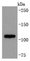
Western blot analysis of ADAM17 on Jurkat cells lysates using anti-ADAM17 antibody at 1/1,000 dilution.ICC/IF
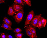
ICC staining ADAM17 in HepG2 cells (red). The nuclear counter stain is DAPI (blue). Cells were fixed in paraformaldehyde, permeabilised with 0.25% Triton X100/PBS.ICC/IF
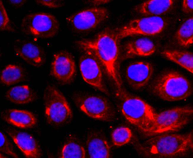
ICC staining ADAM17 in SKOV-3 cells (red). The nuclear counter stain is DAPI (blue). Cells were fixed in paraformaldehyde, permeabilised with 0.25% Triton X100/PBS.ICC/IF
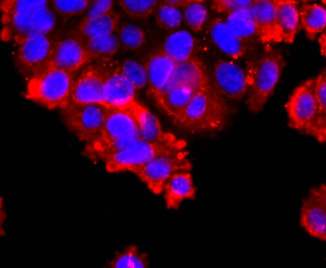
ICC staining ADAM17 in SW480 cells (red). The nuclear counter stain is DAPI (blue). Cells were fixed in paraformaldehyde, permeabilised with 0.25% Triton X100/PBS.FC
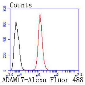
Flow cytometric analysis of Hela cells with ADAM17 antibody at 1/50 dilution (red) compared with an unlabelled control (cells without incubation with primary antibody; black). Alexa Fluor 488-conjugated goat anti rabbit IgG was used as the secondary antibody.IHC
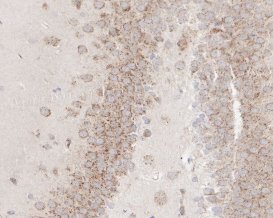
Immunohistochemical analysis of paraffin-embedded rat hippocampus tissue with Rabbit anti-ADAM17 antibody at 1/500 dilution. The section was pre-treated using heat mediated antigen retrieval with Tris-EDTA buffer (pH 9.0) for 20 minutes. The tissues were blocked in 1% BSA for 20 minutes at room temperature, washed with ddH2O and PBS, and then probed with the primary antibody at 1/500 dilution for 1 hour at room temperature. The detection was performed using an HRP conjugated compact polymer system. DAB was used as the chromogen. Tissues were counterstained with hematoxylin and mounted with DPX.IF-F
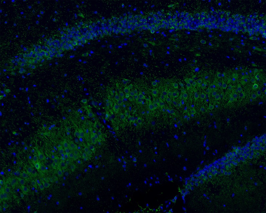
Immunofluorescence analysis of frozen mouse hippocampus tissue labeling ADAM17 with Rabbit anti-ADAM17 antibody. The tissues were blocked in 3% BSA for 30 minutes at room temperature, washed with PBS, and then probed with the primary antibody (green) at 1/100 dilution overnight at 4℃, washed with PBS. Goat Anti-Rabbit IgG H&L (Alexa Fluor® 488) was used as the secondary antibody at 1/200 dilution. Nuclei were counterstained with DAPI (blue). Image acquisition was performed with KFBIO KF-FL-400 Scanner.| Product Name | ADAM17 Recombinant Rabbit Monoclonal Antibody |
|---|---|
| Antibody Type | Primary Antibodies |
| Product description | Tumor necrosis factor β (TNFβ), also known as lymphotoxin, is a pleiotropic cytokine. TNFα, also known as cachetin, is a cytokine that binds to the same receptors, producing an array of effects similar to those of TNFβ. TNFβ and TNFα share 30% amino acid homology and have similar biological activities. TNFβ is produced by activated lymphocytes, including CD4+ T helper cell type 1 lymphocytes, CD8+ lymphocytes and certain B lymphoblastoid cell lines. TNFα is produced by several different cell types, including lymphocytes, neutrophils and macrophages. TNFβ and TNFα can modulate many immune and inflammatory functions while having the ability to inhibit tumor growth. TACE (for TNFα converting enzyme) is a metalloproteinase that cleaves the membrane-bound TNFα precursor to release soluble TNFα. |
| Immunogen | Synthetic peptide within Human ADAM17 aa 757-790 / 824. |
| Clonality | Monoclonal |
|---|---|
| Isotype | IgG |
| Host Species | Recombinant rabbit |
| Tested Applications | FCICC/IFIF-FIHCWB |
| WB:1:1,000-1:2,000 FC:1:50-1:100 ICC:1:50-1:200 IHC:1:500 IF-F:1:100 |
|
| Species Reactivity | HumanMouseRat |
| Concentration | 1mg/ml |
| Purification | Protein A |
| Alternative Names | A disintegrin and metalloproteinase domain 17 (tumor necrosis factor alpha converting enzyme) antibody A disintegrin and metalloproteinase domain 17 antibody ADA17_HUMAN antibody ADAM 17 antibody ADAM metallopeptidase domain 17 antibody ADAM17 antibody ADAM17 protein antibody CD 156b antibody CD156b antibody CD156b antigen antibody CSVP antibody Disintegrin and metalloproteinase domain-containing protein 17 antibody MGC71942 antibody NISBD antibody NISBD1 antibody Snake venom like protease antibody Snake venom-like protease antibody TACE antibody TNF alpha convertase antibody TNF alpha converting enzyme antibody TNF-alpha convertase antibody TNF-alpha-converting enzyme antibody Tumor Necrosis Factor Alpha Converting Enzyme antibody |
|---|---|
| Molecular Weight(MW) | 93 kDa |
| Cellular Localization | Membrane. |
WB
Western blot analysis of ADAM17 on Jurkat cells lysates using anti-ADAM17 antibody at 1/1,000 dilution.
ICC/IF
ICC staining ADAM17 in HepG2 cells (red). The nuclear counter stain is DAPI (blue). Cells were fixed in paraformaldehyde, permeabilised with 0.25% Triton X100/PBS.
ICC/IF
ICC staining ADAM17 in SKOV-3 cells (red). The nuclear counter stain is DAPI (blue). Cells were fixed in paraformaldehyde, permeabilised with 0.25% Triton X100/PBS.
ICC/IF
ICC staining ADAM17 in SW480 cells (red). The nuclear counter stain is DAPI (blue). Cells were fixed in paraformaldehyde, permeabilised with 0.25% Triton X100/PBS.
FC
Flow cytometric analysis of Hela cells with ADAM17 antibody at 1/50 dilution (red) compared with an unlabelled control (cells without incubation with primary antibody; black). Alexa Fluor 488-conjugated goat anti rabbit IgG was used as the secondary antibody.
IHC
Immunohistochemical analysis of paraffin-embedded rat hippocampus tissue with Rabbit anti-ADAM17 antibody at 1/500 dilution. The section was pre-treated using heat mediated antigen retrieval with Tris-EDTA buffer (pH 9.0) for 20 minutes. The tissues were blocked in 1% BSA for 20 minutes at room temperature, washed with ddH2O and PBS, and then probed with the primary antibody at 1/500 dilution for 1 hour at room temperature. The detection was performed using an HRP conjugated compact polymer system. DAB was used as the chromogen. Tissues were counterstained with hematoxylin and mounted with DPX.
IF-F
Immunofluorescence analysis of frozen mouse hippocampus tissue labeling ADAM17 with Rabbit anti-ADAM17 antibody. The tissues were blocked in 3% BSA for 30 minutes at room temperature, washed with PBS, and then probed with the primary antibody (green) at 1/100 dilution overnight at 4℃, washed with PBS. Goat Anti-Rabbit IgG H&L (Alexa Fluor® 488) was used as the secondary antibody at 1/200 dilution. Nuclei were counterstained with DAPI (blue). Image acquisition was performed with KFBIO KF-FL-400 Scanner.| Positive Control | Jurkat, SKOV-3, SW480, HepG2. |
|---|---|
| Application Notes | WB:1:1,000-1:2,000 FC:1:50-1:100 ICC:1:50-1:200 IHC:1:500 IF-F:1:100 |
| Form | Liquid |
|---|---|
| Storage Instructions | Store at +4℃ after thawing. Aliquot store at -20℃ or -80℃. Avoid repeated freeze / thaw cycles. |
| Storage Buffer | 1*TBS (pH7.4), 1%BSA, 40%Glycerol. Preservative: 0.05% Sodium Azide. |


