Application
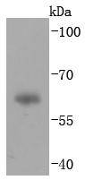
Fig1: Western blot analysis of IgA on human plasma lysates using anti-IgA antibody at 1/1,000 dilution.Application
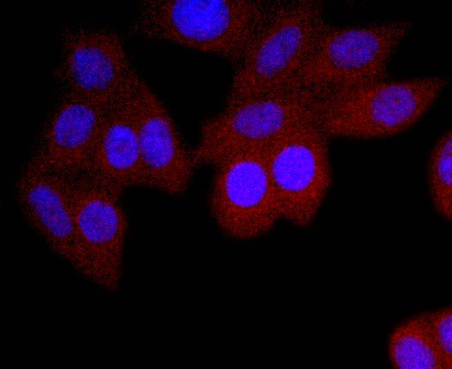
Fig2: ICC staining IgA in HepG2 cells (red). The nuclear counter stain is DAPI (blue). Cells were fixed in paraformaldehyde, permeabilised with 0.25% Triton X100/PBS.Application
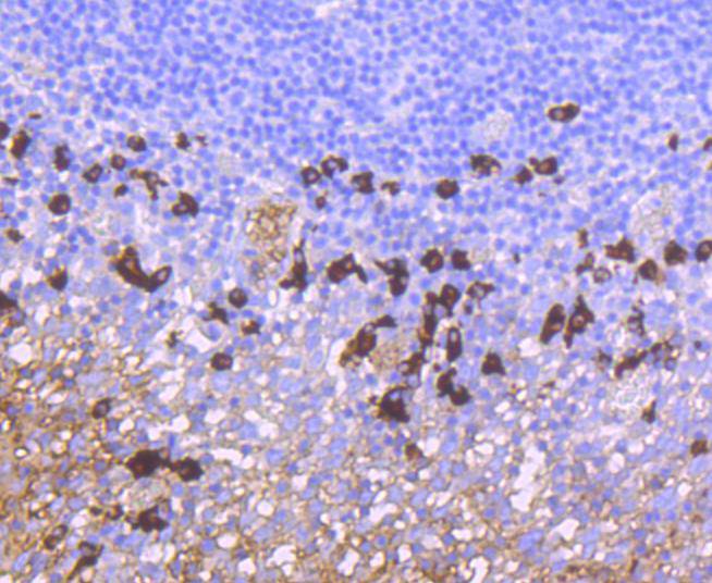
Fig3: Immunohistochemical analysis of paraffin-embedded human tonsil tissue using anti-IgA antibody. Counter stained with hematoxylin.Application
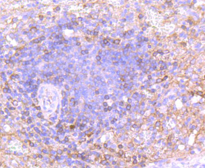
Fig4: Immunohistochemical analysis of paraffin-embedded human spleen tissue using anti-IgA antibody. Counter stained with hematoxylin.| Product Name | IgA [JM10-42] |
|---|---|
| Antibody Type | Primary Antibodies |
| Antigen Alias | Hepatocellular carcinoma-associated protein TB6 antibody Ig alpha 1 chain C region antibody Ig alpha 2 chain C region antibody IGHA antibody IGHA1 antibody IGHA2 antibody Immunoglobulin heavy constant alpha 1 antibody Immunoglobulin heavy constant alpha 2 A2m marker antibody Immunoglobulin heavy constant alpha 2 antibody PIgR antibody Poly-Ig receptor antibody polymeric immunoglobulin receptor antibody polymeric immunoglobulin receptor Secretory component antibody |
| Product description | Immunoglobulins are four-chain, Y-shaped, monomeric structures comprised of two identical heavy chains and two identical light chains held together through interchain disulfide bonds. The chains form two domains, the Fab (antigen binding) fragment and the Fc (constant) fragment. Immunoglobulin A (IgA) is the main protein of the mucosal immune system. It is generated by B cells in gut-associated lymphoid tissues. Daily production of IgA exceeds that of any of the other immunoglobulins. The IgA heavy chain is an α-chain, and the light chains are either κ- or λ- chains. IgA exists mainly in dimers but can also exist as polymers or as monomers. Dimers and polymers contain a joining (J) chain that can be bound by the polymeric immunoglobulin receptor (pIgR) for transportation of the molecule to mucosal surfaces. |
| Immunogen | recombinant protein |
| Clonality | Monoclonal |
|---|---|
| Isotype | IgG |
| Host Species | Recombinant rabbit |
| Tested Applications | WBICC/IFIHCIP |
| WB:1:1,000 ICC:1:50-1:200 IHC:1:50-1:200 IP:1:50-1:100 |
|
| Species Reactivity | Human |
| Concentration | 1mg/ml |
| Alternative Names | Hepatocellular carcinoma-associated protein TB6 antibody Ig alpha 1 chain C region antibody Ig alpha 2 chain C region antibody IGHA antibody IGHA1 antibody IGHA2 antibody Immunoglobulin heavy constant alpha 1 antibody Immunoglobulin heavy constant alpha 2 A2m marker antibody Immunoglobulin heavy constant alpha 2 antibody PIgR antibody Poly-Ig receptor antibody polymeric immunoglobulin receptor antibody polymeric immunoglobulin receptor Secretory component antibody |
|---|---|
| Molecular Weight(MW) | 60 kDa |
| Cellular Localization | Secreted. |
Application
Fig1: Western blot analysis of IgA on human plasma lysates using anti-IgA antibody at 1/1,000 dilution.
Application
Fig2: ICC staining IgA in HepG2 cells (red). The nuclear counter stain is DAPI (blue). Cells were fixed in paraformaldehyde, permeabilised with 0.25% Triton X100/PBS.
Application
Fig3: Immunohistochemical analysis of paraffin-embedded human tonsil tissue using anti-IgA antibody. Counter stained with hematoxylin.
Application
Fig4: Immunohistochemical analysis of paraffin-embedded human spleen tissue using anti-IgA antibody. Counter stained with hematoxylin.| Positive Control | HepG2, human plasma tissue, human tonsil tissue, human spleen tissue. |
|---|---|
| Application Notes | WB:1:1,000 ICC:1:50-1:200 IHC:1:50-1:200 IP:1:50-1:100 |
| Form | Liquid |
|---|---|
| Storage Instructions | Store at +4℃ after thawing. Aliquot store at -20℃ or -80℃. Avoid repeated freeze / thaw cycles. |
| Storage Buffer | 1*TBS (pH7.4), 1%BSA, 40%Glycerol. Preservative: 0.05% Sodium Azide. |


