WB
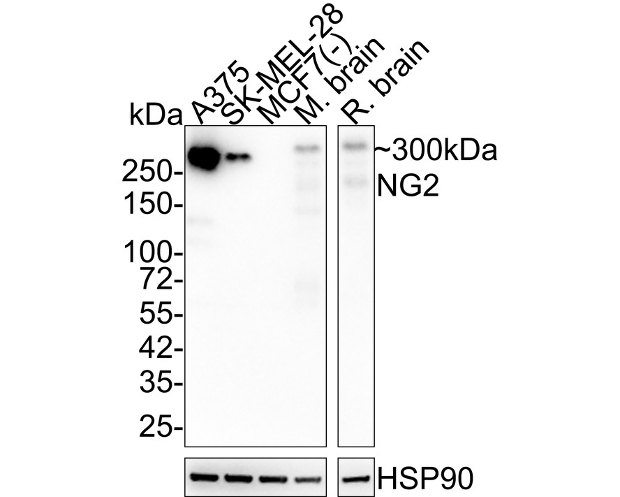
Western blot analysis of NG2 on different lysates with Rabbit anti-NG2 antibody at 1/2,000 dilution. Lane 1: A375 cell lysate (15 µg/Lane), Lane 2: SK-MEL-28 cell lysate (15 µg/Lane), Lane 3: MCF7 cell lysate (negative) (15 µg/Lane), Lane 4: Mouse brain tissue lysate (20 µg/Lane), Lane 5: Rat brain tissue lysate (20 µg/Lane), Exposure time: 2 minutes; 4-20% SDS-PAGE gel. Proteins were transferred to a PVDF membrane and blocked with 5% NFDM/TBST for 1 hour at room temperature. The primary antibody at 1/2,000 dilution was used in 5% NFDM/TBST at 4℃ overnight. Goat Anti-Rabbit IgG - HRP Secondary Antibody at 1/50,000 dilution was used for 1 hour at room temperature.WB
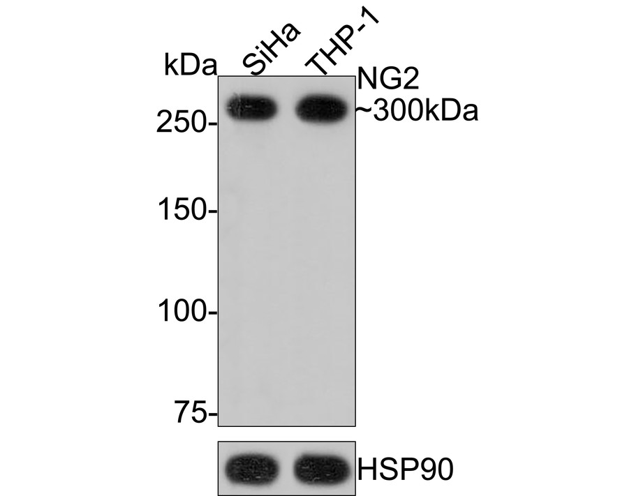
Western blot analysis of NG2 on different lysates with Rabbit anti-NG2 antibody at 1/1,000 dilution. Lane 1: SiHa cell lysate, Lane 2: THP-1 cell lysate, Lysates/proteins at 10 µg/Lane. Exposure time: 3 minutes; 6% SDS-PAGE gel. Proteins were transferred to a PVDF membrane and blocked with 5% NFDM/TBST for 1 hour at room temperature. The primary antibody at 1/1,000 dilution was used in 5% NFDM/TBST at room temperature for 2 hours. Goat Anti-Rabbit IgG - HRP Secondary Antibody at 1:100,000 dilution was used for 1 hour at room temperature.IHC
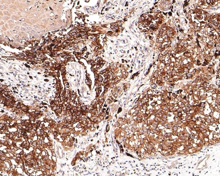
Immunohistochemical analysis of paraffin-embedded human malignant melanoma tissue using anti-NG2 antibody. The section was pre-treated using heat mediated antigen retrieval with Tris-EDTA buffer (pH 9.0) for 20 minutes.The tissues were blocked in 1% BSA for 30 minutes at room temperature, washed with ddH2O and PBS, and then probed with the primary antibody (1/400) for 30 minutes at room temperature. The detection was performed using an HRP conjugated compact polymer system. DAB was used as the chromogen. Tissues were counterstained with hematoxylin and mounted with DPX.IHC
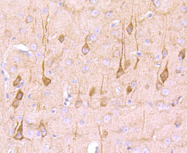
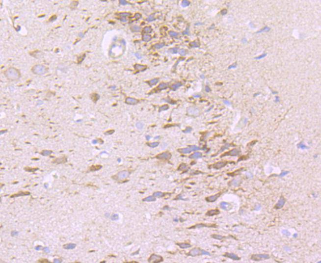
Immunohistochemical analysis of paraffin-embedded mouse brain tissue using anti-NG2 antibody. The section was pre-treated using heat mediated antigen retrieval with Tris-EDTA buffer (pH 8.0-8.4) for 20 minutes.The tissues were blocked in 5% BSA for 30 minutes at room temperature, washed with ddH2O and PBS, and then probed with the primary antibody (1/50) for 30 minutes at room temperature. The detection was performed using an HRP conjugated compact polymer system. DAB was used as the chromogen. Tissues were counterstained with hematoxylin and mounted with DPX.IHC
Immunohistochemical analysis of paraffin-embedded rat brain tissue using anti-NG2 antibody. The section was pre-treated using heat mediated antigen retrieval with Tris-EDTA buffer (pH 8.0-8.4) for 20 minutes.The tissues were blocked in 5% BSA for 30 minutes at room temperature, washed with ddH2O and PBS, and then probed with the primary antibody (1/50) for 30 minutes at room temperature. The detection was performed using an HRP conjugated compact polymer system. DAB was used as the chromogen. Tissues were counterstained with hematoxylin and mounted with DPX.| Product Name | NG2 Recombinant Rabbit Monoclonal Antibody |
|---|---|
| Antibody Type | Primary Antibodies |
| Product description | NG2 (also known as melanoma-associated chondroitin sulfate proteoglycan 4, MCSP, MCSPG, MSK16 and MEL-CSPG) stabilizes cell-substratum interactions during early events of melanoma cell spreading on endothelial basement membranes. NG2 may facilitate primary melanoma progression by enhancing the activation of key signaling pathways important for tumor invasion and growth. Threonine 2256 phosphorylation of rat NG2 (Threonine 2252 phosphorylation of human NG2) leads to redistribution of NG2 on the surface of astrocytomas, polarization of the cell and a significant increase in cell motility. NG2 acts as a co-receptor for spreading and focal contact formation in association with α4 β1 integrin in malignant melanoma cells. NG2 is present on blood vessels throughout the rat embryo. Microvessels within the rat CNS express NG2 on endothelial cells, and outside the CNS, NG2 is present on smooth muscle cells. NG2 is a novel marker for epidermal stem cells that contributes to their patterned distribution by promoting stem cell clustering. |
| Immunogen | Synthetic peptide within Human NG2 aa 2,271-2,314 / 2,322. |
| Clonality | Monoclonal |
|---|---|
| Isotype | IgG |
| Host Species | Recombinant rabbit |
| Tested Applications | IHCWB |
| WB:1:1000-1:2000 IHC:1:50-1:400 |
|
| Species Reactivity | HumanMouseRat |
| Concentration | 1mg/ml |
| Purification | Protein A |
| Alternative Names | 4732461B14Rik antibody AN2 antibody AN2 proteoglycan antibody Chondroitin sulfate proteoglycan 4 (melanoma-associated) antibody Chondroitin sulfate proteoglycan 4 antibody Chondroitin sulfate proteoglycan NG2 antibody Cspg4 antibody Cspg4 chondroitin sulfate proteoglycan 4 antibody CSPG4_HUMAN antibody HMW-MAA antibody HSN tumor-specific antigen antibody Kiaa4232 antibody MCSP antibody MCSPG antibody MEL-CSPG antibody Melanoma chondroitin sulfate proteoglycan antibody Melanoma-associated chondroitin sulfate proteoglycan antibody MELCSPG antibody MSK16 antibody NG2 antibody |
|---|---|
| Molecular Weight(MW) | 251 kDa(Observed band size: 300 kDa) |
| Cellular Localization | Cell membrane, Apical cell membrane, Cell projection, Cell surface. |
| SwissProt ID | Q6UVK1 |
|---|
WB
Western blot analysis of NG2 on different lysates with Rabbit anti-NG2 antibody at 1/2,000 dilution. Lane 1: A375 cell lysate (15 µg/Lane), Lane 2: SK-MEL-28 cell lysate (15 µg/Lane), Lane 3: MCF7 cell lysate (negative) (15 µg/Lane), Lane 4: Mouse brain tissue lysate (20 µg/Lane), Lane 5: Rat brain tissue lysate (20 µg/Lane), Exposure time: 2 minutes; 4-20% SDS-PAGE gel. Proteins were transferred to a PVDF membrane and blocked with 5% NFDM/TBST for 1 hour at room temperature. The primary antibody at 1/2,000 dilution was used in 5% NFDM/TBST at 4℃ overnight. Goat Anti-Rabbit IgG - HRP Secondary Antibody at 1/50,000 dilution was used for 1 hour at room temperature.
WB
Western blot analysis of NG2 on different lysates with Rabbit anti-NG2 antibody at 1/1,000 dilution. Lane 1: SiHa cell lysate, Lane 2: THP-1 cell lysate, Lysates/proteins at 10 µg/Lane. Exposure time: 3 minutes; 6% SDS-PAGE gel. Proteins were transferred to a PVDF membrane and blocked with 5% NFDM/TBST for 1 hour at room temperature. The primary antibody at 1/1,000 dilution was used in 5% NFDM/TBST at room temperature for 2 hours. Goat Anti-Rabbit IgG - HRP Secondary Antibody at 1:100,000 dilution was used for 1 hour at room temperature.
IHC
Immunohistochemical analysis of paraffin-embedded human malignant melanoma tissue using anti-NG2 antibody. The section was pre-treated using heat mediated antigen retrieval with Tris-EDTA buffer (pH 9.0) for 20 minutes.The tissues were blocked in 1% BSA for 30 minutes at room temperature, washed with ddH2O and PBS, and then probed with the primary antibody (1/400) for 30 minutes at room temperature. The detection was performed using an HRP conjugated compact polymer system. DAB was used as the chromogen. Tissues were counterstained with hematoxylin and mounted with DPX.
IHC
Immunohistochemical analysis of paraffin-embedded mouse brain tissue using anti-NG2 antibody. The section was pre-treated using heat mediated antigen retrieval with Tris-EDTA buffer (pH 8.0-8.4) for 20 minutes.The tissues were blocked in 5% BSA for 30 minutes at room temperature, washed with ddH2O and PBS, and then probed with the primary antibody (1/50) for 30 minutes at room temperature. The detection was performed using an HRP conjugated compact polymer system. DAB was used as the chromogen. Tissues were counterstained with hematoxylin and mounted with DPX.
IHC
Immunohistochemical analysis of paraffin-embedded rat brain tissue using anti-NG2 antibody. The section was pre-treated using heat mediated antigen retrieval with Tris-EDTA buffer (pH 8.0-8.4) for 20 minutes.The tissues were blocked in 5% BSA for 30 minutes at room temperature, washed with ddH2O and PBS, and then probed with the primary antibody (1/50) for 30 minutes at room temperature. The detection was performed using an HRP conjugated compact polymer system. DAB was used as the chromogen. Tissues were counterstained with hematoxylin and mounted with DPX.| Application Notes | WB:1:1000-1:2000 IHC:1:50-1:400 |
|---|
| Form | Liquid |
|---|---|
| Storage Instructions | Store at +4℃ after thawing. Aliquot store at -20℃ or -80℃. Avoid repeated freeze / thaw cycles. |
| Storage Buffer | 1*TBS (pH7.4), 1%BSA, 40%Glycerol. Preservative: 0.05% Sodium Azide. |


