Application
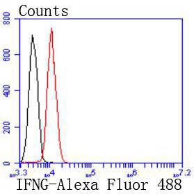
Fig4: Flow cytometric analysis of Hela cells with Interferon gamma antibody at 1/50 dilution (red) compared with an unlabelled control (cells without incubation with primary antibody; black). Alexa Fluor 488-conjugated goat anti rabbit IgG was used as the secondary antibody.Application
Fig4: Flow cytometric analysis of Hela cells with Interferon gamma antibody at 1/50 dilution (red) compared with an unlabelled control (cells without incubation with primary antibody; black). Alexa Fluor 488-conjugated goat anti rabbit IgG was used as the secondary antibody.Application
Fig4: Flow cytometric analysis of Hela cells with Interferon gamma antibody at 1/50 dilution (red) compared with an unlabelled control (cells without incubation with primary antibody; black). Alexa Fluor 488-conjugated goat anti rabbit IgG was used as the secondary antibody.Application
Fig4: Flow cytometric analysis of Hela cells with Interferon gamma antibody at 1/50 dilution (red) compared with an unlabelled control (cells without incubation with primary antibody; black). Alexa Fluor 488-conjugated goat anti rabbit IgG was used as the secondary antibody.| Product Name | Interferon gamma [JM10-10] |
|---|---|
| Antibody Type | Primary Antibodies |
| Antigen Alias | IFG antibody IFI antibody IFN gamma antibody IFN, immune antibody IFN-gamma antibody IFNG antibody IFNG_HUMAN antibody Immune interferon antibody Interferon gamma antibody |
| Product description | Interferon (IFN)-γ is an antiviral and antiparasitic agent produced by CD4+/CD8+ lymphocytes and natural killer cells that undergo activation by antigens, mitogens or alloantigens. IFN-γ production modulates T cell growth and differentiation and inhibits the growth of B cells. Synthesis of IFN-γ is inducible by IL-2, FGF and EGF. The active form of IFN-γ is a homodimer with each subunit containing six helices. The dimeric structure of human IFN-γ is stabilized by non-covalent interactions through the interface of the helices. IFN-γ translated precursor is 166 amino acids, including the 23 amino acid secretory sequence. Multiple forms exist due to variable glycosylation and under non-denaturing conditions due to dimers and tetramers. |
| Immunogen | recombinant protein |
| Clonality | Monoclonal |
|---|---|
| Isotype | IgG |
| Host Species | Recombinant rabbit |
| Tested Applications | WBIHCFC |
| WB:1:500-1:1,000 FC:1:50-1:100 IHC:1:50-1:100 |
|
| Species Reactivity | HumanMouseRat |
| Concentration | 1mg/ml |
| Alternative Names | IFG antibody
IFI antibody
IFN gamma antibody
IFN immune antibody IFN-gamma antibody IFNG antibody IFNG_HUMAN antibody Immune interferon antibody Interferon gamma antibody |
|---|---|
| Molecular Weight(MW) | 19 kDa |
| Cellular Localization | Secreted. |
Application
Fig4: Flow cytometric analysis of Hela cells with Interferon gamma antibody at 1/50 dilution (red) compared with an unlabelled control (cells without incubation with primary antibody; black). Alexa Fluor 488-conjugated goat anti rabbit IgG was used as the secondary antibody.
Application
Fig4: Flow cytometric analysis of Hela cells with Interferon gamma antibody at 1/50 dilution (red) compared with an unlabelled control (cells without incubation with primary antibody; black). Alexa Fluor 488-conjugated goat anti rabbit IgG was used as the secondary antibody.
Application
Fig4: Flow cytometric analysis of Hela cells with Interferon gamma antibody at 1/50 dilution (red) compared with an unlabelled control (cells without incubation with primary antibody; black). Alexa Fluor 488-conjugated goat anti rabbit IgG was used as the secondary antibody.
Application
Fig4: Flow cytometric analysis of Hela cells with Interferon gamma antibody at 1/50 dilution (red) compared with an unlabelled control (cells without incubation with primary antibody; black). Alexa Fluor 488-conjugated goat anti rabbit IgG was used as the secondary antibody.| Positive Control | Human tonsil tissue, human spleen tissue, mouse spleen tissue. |
|---|---|
| Application Notes | WB:1:500-1:1,000 FC:1:50-1:100 IHC:1:50-1:100 |
| Form | Liquid |
|---|---|
| Storage Instructions | Store at +4℃ after thawing. Aliquot store at -20℃ or -80℃. Avoid repeated freeze / thaw cycles. |
| Storage Buffer | 1*TBS (pH7.4), 1%BSA, 40%Glycerol. Preservative: 0.05% Sodium Azide. |


