WB
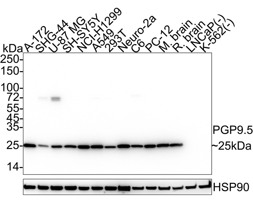
Western blot analysis of PGP9.5 on different lysates with Rabbit anti-PGP9.5 antibody at 1/2,000 dilution. Lane 1: A-172 cell lysate (20 µg/Lane), Lane 2: SHG-44 cell lysate (20 µg/Lane), Lane 3: U-87 MG cell lysate (20 µg/Lane), Lane 4: SH-SY5Y cell lysate (20 µg/Lane), Lane 5: NCI-H1299 cell lysate (20 µg/Lane), Lane 6: A549 cell lysate (20 µg/Lane), Lane 7: 293T cell lysate (20 µg/Lane), Lane 8: Neuro-2a cell lysate (20 µg/Lane), Lane 9: C6 cell lysate (20 µg/Lane), Lane 10: PC-12 cell lysate (20 µg/Lane), Lane 11: Mouse brain tissue lysate (30 µg/Lane), Lane 12: Rat brain tissue lysate (30 µg/Lane), Lane 13: LNCaP cell lysate (negative) (20 µg/Lane), Lane 14: K-562 cell lysate (negative) (20 µg/Lane), Exposure time: 7 seconds; 4-20% SDS-PAGE gel. Proteins were transferred to a PVDF membrane and blocked with 5% NFDM/TBST for 1 hour at room temperature. The primary antibody at 1/2,000 dilution was used in 5% NFDM/TBST at 4℃ overnight. Goat Anti-Rabbit IgG - HRP Secondary Antibody at 1/50,000 dilution was used for 1 hour at room temperature.IHC
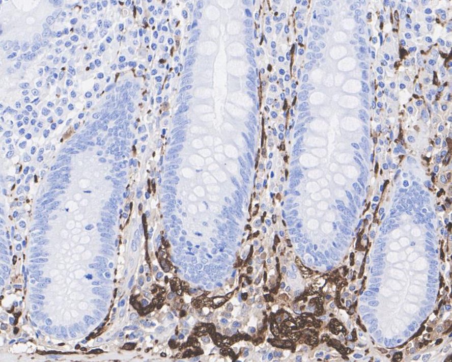
Immunohistochemical analysis of paraffin-embedded human appendix tissue with Rabbit anti-PGP9.5 antibody at 1/5,000 dilution. The section was pre-treated using heat mediated antigen retrieval with Tris-EDTA buffer (pH 9.0) for 20 minutes. The tissues were blocked in 1% BSA for 20 minutes at room temperature, washed with ddH2O and PBS, and then probed with the primary antibody at 1/5,000 dilution for 1 hour at room temperature. The detection was performed using an HRP conjugated compact polymer system. DAB was used as the chromogen. Tissues were counterstained with hematoxylin and mounted with DPX.IHC
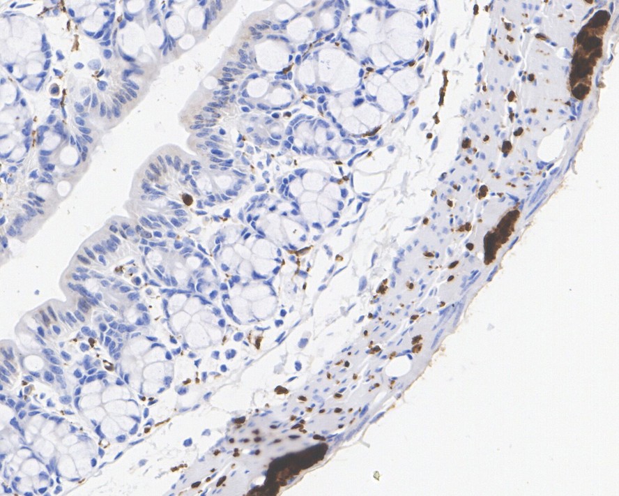
Immunohistochemical analysis of paraffin-embedded mouse colon tissue with Rabbit anti-PGP9.5 antibody at 1/5,000 dilution. The section was pre-treated using heat mediated antigen retrieval with Tris-EDTA buffer (pH 9.0) for 20 minutes. The tissues were blocked in 1% BSA for 20 minutes at room temperature, washed with ddH2O and PBS, and then probed with the primary antibody at 1/5,000 dilution for 1 hour at room temperature. The detection was performed using an HRP conjugated compact polymer system. DAB was used as the chromogen. Tissues were counterstained with hematoxylin and mounted with DPX.IHC
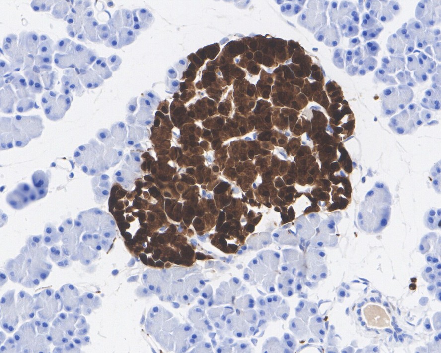
Immunohistochemical analysis of paraffin-embedded rat pancreas tissue with Rabbit anti-PGP9.5 antibody at 1/5,000 dilution. The section was pre-treated using heat mediated antigen retrieval with Tris-EDTA buffer (pH 9.0) for 20 minutes. The tissues were blocked in 1% BSA for 20 minutes at room temperature, washed with ddH2O and PBS, and then probed with the primary antibody at 1/5,000 dilution for 1 hour at room temperature. The detection was performed using an HRP conjugated compact polymer system. DAB was used as the chromogen. Tissues were counterstained with hematoxylin and mounted with DPX.ICC/IF
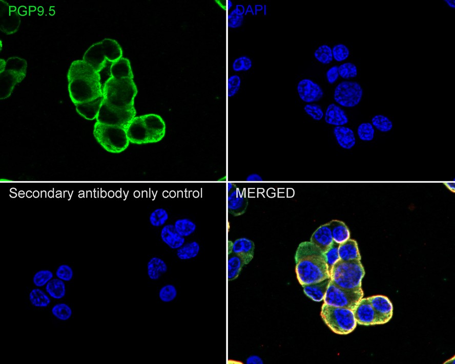
Immunocytochemistry analysis of PC-12 cells labeling PGP9.5 with Rabbit anti-PGP9.5 antibody at 1/100 dilution. Cells were fixed in 4% paraformaldehyde for 20 minutes at room temperature, permeabilized with 0.1% Triton X-100 in PBS for 5 minutes at room temperature, then blocked with 1% BSA in 10% negative goat serum for 1 hour at room temperature. Cells were then incubated with Rabbit anti-PGP9.5 antibody at 1/100 dilution in 1% BSA in PBST overnight at 4 ℃. Goat Anti-Rabbit IgG H&L (488) was used as the secondary antibody at 1/1,000 dilution. PBS instead of the primary antibody was used as the secondary antibody only control. Nuclear DNA was labelled in blue with DAPI. Beta tubulin (red) was stained at 1/100 dilution overnight at +4℃. Goat Anti-Mouse IgG H&L (594) was used as the secondary antibody at 1/1,000 dilution.IF-F
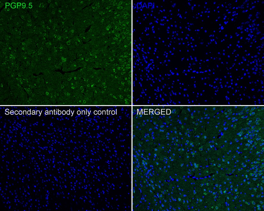
Species: Mouse, Site: Cerebral cortex, Sample: Frozen section, Antibody concentration: 1/200, Antigen retrieval: The section was pre-treated using heat mediated antigen retrieval with sodium citrate buffer (pH 6.0) for about 2 minutes in microwave oven.IF-F
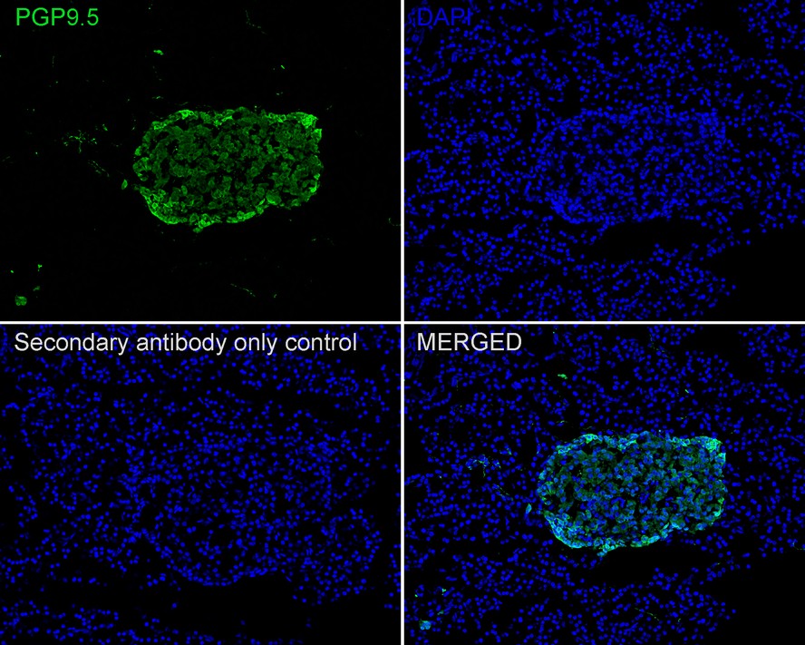
Species: Mouse, Site: Pancreas, Sample: Frozen section, Antibody concentration: 1/200, Antigen retrieval: The section was pre-treated using heat mediated antigen retrieval with sodium citrate buffer (pH 6.0) for about 2 minutes in microwave oven.FC
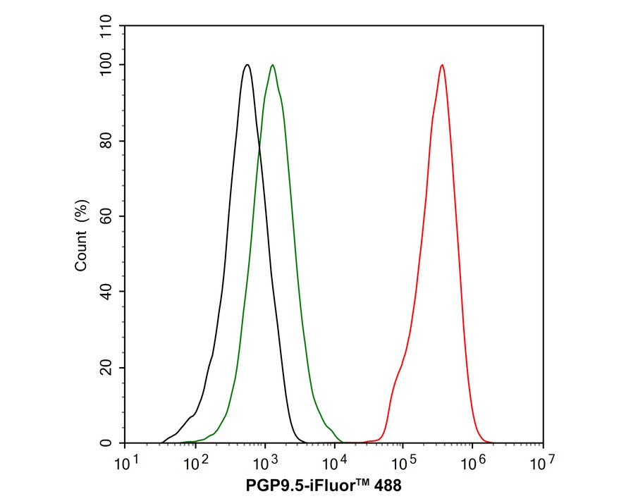
Flow cytometric analysis of SH-SY5Y cells labeling PGP9.5. Cells were fixed and permeabilized. Then stained with the primary antibody (1μg/mL) (red) compared with Rabbit IgG Isotype Control (green). After incubation of the primary antibody at +4℃ for an hour, the cells were stained with a iFluor™ 488 conjugate-Goat anti-Rabbit IgG Secondary antibody at 1/1,000 dilution for 30 minutes at +4℃. Unlabelled sample was used as a control (cells without incubation with primary antibody; black).| Product Name | PGP9.5 Recombinant Rabbit Monoclonal Antibody |
|---|---|
| Antibody Type | Primary Antibodies |
| Product description | UCH-L1 (ubiquitin C-terminal hydrolase) is a member of a gene family whose products hydrolyze small C-terminal adducts of ubiquitin to generate the ubiquitin monomer. Expression of UCH-L1 is highly specific to neurons and to cells of the diffuse neuroendocrine system and their tumors. UCH-L1 is expressed in brain neurons. Examination of specific brain regions reveals expression in all areas tested, particularly in the substantia nigra. UCH-L1 represents 1-2% of total soluble brain protein. Its occurrence in Lewy bodies and its function in the proteasome pathway make it a compelling candidate gene in Parkinson disease. The gene which encodes UCH-L1 maps to human chromosome 4p13. The 230 amino acid human UCH-L3 protein is 54% identical to that of UCH-L1. UCH-L3 is the predominant thiol protease and has high-affinity binding sites for ubiquitin. |
| Immunogen | Synthetic peptide within Human PGP95 aa 191-223 / 223. |
| Clonality | Monoclonal |
|---|---|
| Isotype | IgG |
| Host Species | Recombinant rabbit |
| Tested Applications | FCICC/IFIF-FIHCWB |
| WB:1:2000-1:5000 IHC:1:1000-1:5000 ICC/IF:1:100-1:500 IF-F:1:200-1:500 FC:1:1000 |
|
| Species Reactivity | HumanMouseRat |
| Concentration | 1mg/ml |
| Purification | Protein A |
| Alternative Names | Epididymis luminal protein 117 antibody Epididymis secretory protein Li 53 antibody HEL 117 antibody HEL S 53 antibody NDGOA antibody Neuron cytoplasmic protein 9.5 antibody OTTHUMP00000218137 antibody OTTHUMP00000218139 antibody OTTHUMP00000218140 antibody OTTHUMP00000218141 antibody Park 5 antibody PARK5 antibody PGP 9.5 antibody PGP9.5 antibody PGP95 antibody Protein gene product 9.5 antibody Ubiquitin C terminal esterase L1 antibody Ubiquitin C terminal hydrolase antibody Ubiquitin C terminal hydrolase L1 antibody Ubiquitin carboxyl terminal esterase L1 antibody Ubiquitin carboxyl terminal hydrolase isozyme L1 antibody Ubiquitin carboxyl-terminal hydrolase isozyme L1 antibody Ubiquitin thioesterase L1 antibody Ubiquitin thiolesterase antibody Ubiquitin thiolesterase L1 antibody UCH-L1 antibody UCHL1 antibody UCHL1_HUMAN antibody |
|---|---|
| Molecular Weight(MW) | 25 kDa |
| Cellular Localization | Cytoplasm, Endoplasmic reticulum membrane. |
WB
Western blot analysis of PGP9.5 on different lysates with Rabbit anti-PGP9.5 antibody at 1/2,000 dilution. Lane 1: A-172 cell lysate (20 µg/Lane), Lane 2: SHG-44 cell lysate (20 µg/Lane), Lane 3: U-87 MG cell lysate (20 µg/Lane), Lane 4: SH-SY5Y cell lysate (20 µg/Lane), Lane 5: NCI-H1299 cell lysate (20 µg/Lane), Lane 6: A549 cell lysate (20 µg/Lane), Lane 7: 293T cell lysate (20 µg/Lane), Lane 8: Neuro-2a cell lysate (20 µg/Lane), Lane 9: C6 cell lysate (20 µg/Lane), Lane 10: PC-12 cell lysate (20 µg/Lane), Lane 11: Mouse brain tissue lysate (30 µg/Lane), Lane 12: Rat brain tissue lysate (30 µg/Lane), Lane 13: LNCaP cell lysate (negative) (20 µg/Lane), Lane 14: K-562 cell lysate (negative) (20 µg/Lane), Exposure time: 7 seconds; 4-20% SDS-PAGE gel. Proteins were transferred to a PVDF membrane and blocked with 5% NFDM/TBST for 1 hour at room temperature. The primary antibody at 1/2,000 dilution was used in 5% NFDM/TBST at 4℃ overnight. Goat Anti-Rabbit IgG - HRP Secondary Antibody at 1/50,000 dilution was used for 1 hour at room temperature.
IHC
Immunohistochemical analysis of paraffin-embedded human appendix tissue with Rabbit anti-PGP9.5 antibody at 1/5,000 dilution. The section was pre-treated using heat mediated antigen retrieval with Tris-EDTA buffer (pH 9.0) for 20 minutes. The tissues were blocked in 1% BSA for 20 minutes at room temperature, washed with ddH2O and PBS, and then probed with the primary antibody at 1/5,000 dilution for 1 hour at room temperature. The detection was performed using an HRP conjugated compact polymer system. DAB was used as the chromogen. Tissues were counterstained with hematoxylin and mounted with DPX.
IHC
Immunohistochemical analysis of paraffin-embedded mouse colon tissue with Rabbit anti-PGP9.5 antibody at 1/5,000 dilution. The section was pre-treated using heat mediated antigen retrieval with Tris-EDTA buffer (pH 9.0) for 20 minutes. The tissues were blocked in 1% BSA for 20 minutes at room temperature, washed with ddH2O and PBS, and then probed with the primary antibody at 1/5,000 dilution for 1 hour at room temperature. The detection was performed using an HRP conjugated compact polymer system. DAB was used as the chromogen. Tissues were counterstained with hematoxylin and mounted with DPX.
IHC
Immunohistochemical analysis of paraffin-embedded rat pancreas tissue with Rabbit anti-PGP9.5 antibody at 1/5,000 dilution. The section was pre-treated using heat mediated antigen retrieval with Tris-EDTA buffer (pH 9.0) for 20 minutes. The tissues were blocked in 1% BSA for 20 minutes at room temperature, washed with ddH2O and PBS, and then probed with the primary antibody at 1/5,000 dilution for 1 hour at room temperature. The detection was performed using an HRP conjugated compact polymer system. DAB was used as the chromogen. Tissues were counterstained with hematoxylin and mounted with DPX.
ICC/IF
Immunocytochemistry analysis of PC-12 cells labeling PGP9.5 with Rabbit anti-PGP9.5 antibody at 1/100 dilution. Cells were fixed in 4% paraformaldehyde for 20 minutes at room temperature, permeabilized with 0.1% Triton X-100 in PBS for 5 minutes at room temperature, then blocked with 1% BSA in 10% negative goat serum for 1 hour at room temperature. Cells were then incubated with Rabbit anti-PGP9.5 antibody at 1/100 dilution in 1% BSA in PBST overnight at 4 ℃. Goat Anti-Rabbit IgG H&L (488) was used as the secondary antibody at 1/1,000 dilution. PBS instead of the primary antibody was used as the secondary antibody only control. Nuclear DNA was labelled in blue with DAPI. Beta tubulin (red) was stained at 1/100 dilution overnight at +4℃. Goat Anti-Mouse IgG H&L (594) was used as the secondary antibody at 1/1,000 dilution.
IF-F
Species: Mouse, Site: Cerebral cortex, Sample: Frozen section, Antibody concentration: 1/200, Antigen retrieval: The section was pre-treated using heat mediated antigen retrieval with sodium citrate buffer (pH 6.0) for about 2 minutes in microwave oven.
IF-F
Species: Mouse, Site: Pancreas, Sample: Frozen section, Antibody concentration: 1/200, Antigen retrieval: The section was pre-treated using heat mediated antigen retrieval with sodium citrate buffer (pH 6.0) for about 2 minutes in microwave oven.
FC
Flow cytometric analysis of SH-SY5Y cells labeling PGP9.5. Cells were fixed and permeabilized. Then stained with the primary antibody (1μg/mL) (red) compared with Rabbit IgG Isotype Control (green). After incubation of the primary antibody at +4℃ for an hour, the cells were stained with a iFluor™ 488 conjugate-Goat anti-Rabbit IgG Secondary antibody at 1/1,000 dilution for 30 minutes at +4℃. Unlabelled sample was used as a control (cells without incubation with primary antibody; black).| Application Notes | WB:1:2000-1:5000 IHC:1:1000-1:5000 ICC/IF:1:100-1:500 IF-F:1:200-1:500 FC:1:1000 |
|---|
| Form | Liquid |
|---|---|
| Storage Instructions | Store at +4℃ after thawing. Aliquot store at -20℃ or -80℃. Avoid repeated freeze / thaw cycles. |
| Storage Buffer | 1*TBS (pH7.4), 1%BSA, 40%Glycerol. Preservative: 0.05% Sodium Azide. |


