Application
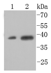
Fig1: Western blot analysis of XBP1 on different cell lysates using anti-XBP1 antibody at 1/1,000 dilution. Positive control: Lane 1:HepG2 Lane 2: 293TApplication
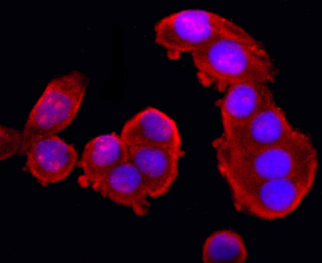
Fig2: ICC staining XBP1 in Hela cells (red). The nuclear counter stain is DAPI (blue). Cells were fixed in paraformaldehyde, permeabilised with 0.25% Triton X100/PBS.Application
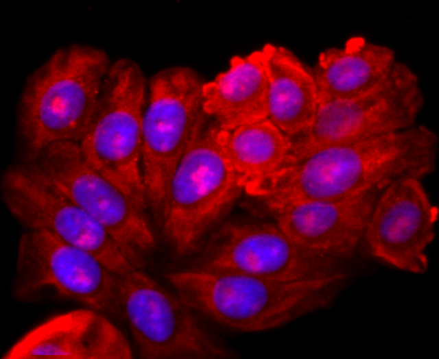
Fig3: ICC staining XBP1 in HepG2 cells (red). The nuclear counter stain is DAPI (blue). Cells were fixed in paraformaldehyde, permeabilised with 0.25% Triton X100/PBS.Application
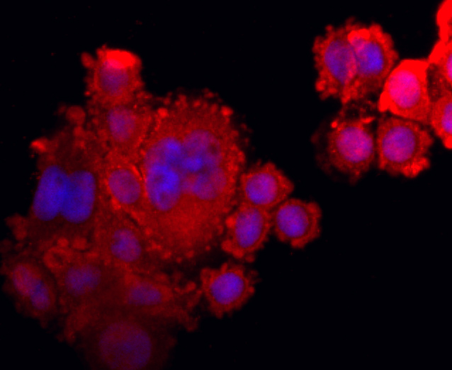
Fig4: ICC staining XBP1 in SW480 cells (red). The nuclear counter stain is DAPI (blue). Cells were fixed in paraformaldehyde, permeabilised with 0.25% Triton X100/PBS.Application
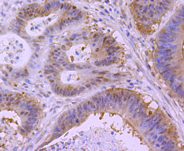
Fig5: Immunohistochemical analysis of paraffin-embedded human colon cancer tissue using anti-XBP1 antibody. Counter stained with hematoxylin.Application
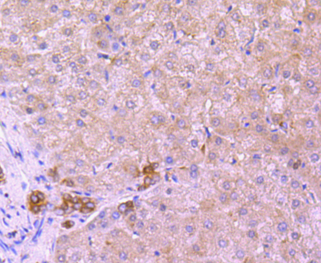
Fig6: Immunohistochemical analysis of paraffin-embedded human liver tissue using anti-XBP1 antibody. Counter stained with hematoxylin.Application
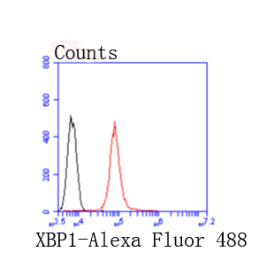
Fig7: Flow cytometric analysis of 293T cells with XBP1 antibody at 1/50 dilution (red) compared with an unlabelled control (cells without incubation with primary antibody; black). Alexa Fluor 488-conjugated goat anti rabbit IgG was used as the secondary antibody.| Product Name | XBP1 [JM10-62] |
|---|---|
| Antibody Type | Primary Antibodies |
| Antigen Alias | Tax responsive element binding protein 5 antibody Tax-responsive element-binding protein 5 antibody TREB5 antibody X box binding protein 1 antibody X box binding protein 2 antibody X-box-binding protein 1 antibody XBP 1 antibody XBP-1 antibody XBP1 antibody XBP1_HUMAN antibody XBP2 antibody |
| Product description | The X-box binding protein-1 (XBP-1 or hXBP-1), also designated tax-responsive element-binding protein 5 (TREB5) in mouse and human, or hepatocarcinogenesis-related transcription factor (HTF) in rat, belongs to the basic region/leucine zipper (bZIP) family of transcription factors. XBP-1 was first characterized as a protein that binds to the HLA-DRα promoter in B cells. XBP-1 recognizes the cAMP responsive element (CRE) in enhancers of human T cell leukemia virus and major histocompatibility complex class II genes and activates transcription of these genes. It is expressed at high levels in developing bone and its levels are modulated during osteoblast development, suggesting a role in regulation of expression of osteoblast-specific genes. In addition to binding to CRE sequences, XBP-1 has been shown to bind to TPA response elements (TREs). |
| Immunogen | recombinant protein |
| Clonality | Monoclonal |
|---|---|
| Isotype | IgG |
| Host Species | Recombinant rabbit |
| Tested Applications | WBIHCICC/IFFC |
| WB:1:500-1:1,000 ICC:1:100-1:500 IHC:1:50-1:200 FC:1:50-1:100 |
|
| Species Reactivity | Human |
| Concentration | 1mg/ml |
| Alternative Names | Tax responsive element binding protein 5 antibody Tax-responsive element-binding protein 5 antibody TREB5 antibody X box binding protein 1 antibody X box binding protein 2 antibody X-box-binding protein 1 antibody XBP 1 antibody XBP-1 antibody XBP1 antibody XBP1_HUMAN antibody XBP2 antibody |
|---|---|
| Molecular Weight(MW) | 40 kDa |
| Cellular Localization | Nucleus, Chromosome, Chromosome. |
Application
Fig1: Western blot analysis of XBP1 on different cell lysates using anti-XBP1 antibody at 1/1,000 dilution. Positive control: Lane 1:HepG2 Lane 2: 293T
Application
Fig2: ICC staining XBP1 in Hela cells (red). The nuclear counter stain is DAPI (blue). Cells were fixed in paraformaldehyde, permeabilised with 0.25% Triton X100/PBS.
Application
Fig3: ICC staining XBP1 in HepG2 cells (red). The nuclear counter stain is DAPI (blue). Cells were fixed in paraformaldehyde, permeabilised with 0.25% Triton X100/PBS.
Application
Fig4: ICC staining XBP1 in SW480 cells (red). The nuclear counter stain is DAPI (blue). Cells were fixed in paraformaldehyde, permeabilised with 0.25% Triton X100/PBS.
Application
Fig5: Immunohistochemical analysis of paraffin-embedded human colon cancer tissue using anti-XBP1 antibody. Counter stained with hematoxylin.
Application
Fig6: Immunohistochemical analysis of paraffin-embedded human liver tissue using anti-XBP1 antibody. Counter stained with hematoxylin.
Application
Fig7: Flow cytometric analysis of 293T cells with XBP1 antibody at 1/50 dilution (red) compared with an unlabelled control (cells without incubation with primary antibody; black). Alexa Fluor 488-conjugated goat anti rabbit IgG was used as the secondary antibody.| Positive Control | Hela, 293T, HepG2, SW480, human liver tissue, human colon cancer tissue. |
|---|---|
| Application Notes | WB:1:500-1:1,000 ICC:1:100-1:500 IHC:1:50-1:200 FC:1:50-1:100 |
| Form | Liquid |
|---|---|
| Storage Instructions | Store at +4℃ after thawing. Aliquot store at -20℃ or -80℃. Avoid repeated freeze / thaw cycles. |
| Storage Buffer | 1*TBS (pH7.4), 1%BSA, 40%Glycerol. Preservative: 0.05% Sodium Azide. |


