Application
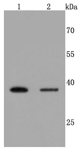
Fig1: Western blot analysis of Haptoglobin on different cells lysates using anti-Haptoglobin antibody at 1/500 dilution. Positive control: Lane 1: Hela Lane 2: HepG2Application
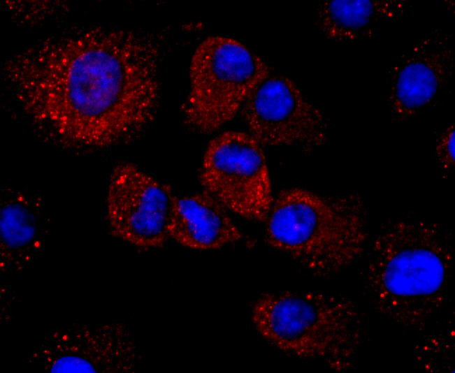
Fig2: ICC staining Haptoglobin in A549 cells (red). The nuclear counter stain is DAPI (blue). Cells were fixed in paraformaldehyde, permeabilised with 0.25% Triton X100/PBS.Application
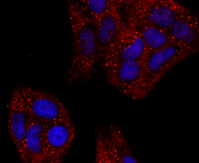
Fig3: ICC staining Haptoglobin in Hela cells (red). The nuclear counter stain is DAPI (blue). Cells were fixed in paraformaldehyde, permeabilised with 0.25% Triton X100/PBS.Application
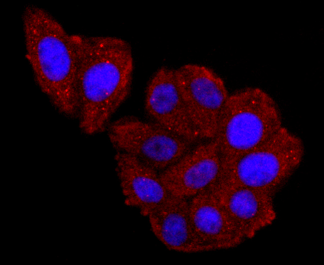
Fig4:ICC staining Haptoglobin in HepG2 cells (red). The nuclear counter stain is DAPI (blue). Cells were fixed in paraformaldehyde, permeabilised with 0.25% Triton X100/PBS.Application
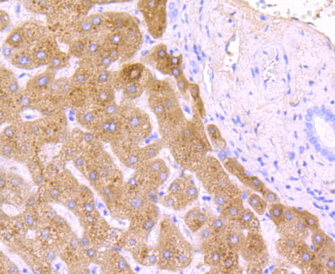
Fig5: Immunohistochemical analysis of paraffin-embedded human liver tissue using anti-Haptoglobin antibody. Counter stained with hematoxylin.Application
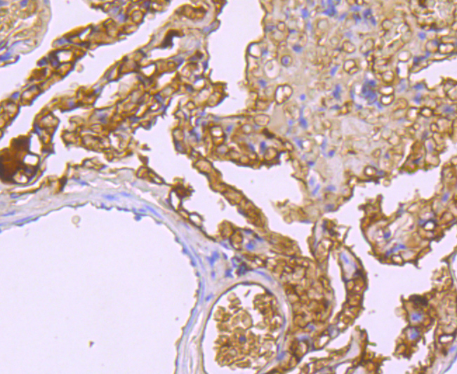
Fig6: Immunohistochemical analysis of paraffin-embedded human lung tissue using anti-Haptoglobin antibody. Counter stained with hematoxylin.Application
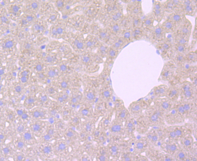
Fig7: Immunohistochemical analysis of paraffin-embedded mouse liver tissue using anti-Haptoglobin antibody. Counter stained with hematoxylin.Application
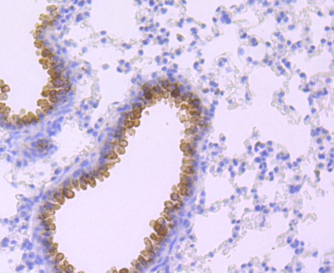
Fig8: Immunohistochemical analysis of paraffin-embedded mouse lung tissue using anti-Haptoglobin antibody. Counter stained with hematoxylin.| Product Name | Haptoglobin [JM10-79] |
|---|---|
| Antibody Type | Primary Antibodies |
| Antigen Alias | Binding peptide antibody BP antibody Haptoglobin alpha chain antibody Haptoglobin alpha(1S) beta antibody Haptoglobin alpha(2FS) beta antibody Haptoglobin beta chain antibody Haptoglobin, alpha polypeptide antibody Haptoglobin, beta polypeptide antibody HP antibody HP2 ALPHA2 antibody HP2ALPHA2 antibody HPA1S antibody HPT antibody HPT_HUMAN antibody MGC111141 antibody Zonulin antibody |
| Product description | Haptoglobin (Hp) is a blood plasma protein that functions to bind free Hemoglobin that has been released from erythrocytes, thereby inhibiting its oxidative activity. During this process, Haptoglobin sequesters the iron within Hemoglobin, preventing iron-utilizing bacteria from benefitting from hemolysis. This function suggests that Haptoglobin concentrations may increase in response to inflammation. The resulting Haptoglobin-Hemoglobin complex is then removed by the reticulo-endothelial system. Due to cleavage of a common precursor protein during protein synthesis, Haptoglobin consists of two α and two β chains, connected by disulfide bridges. In human, Haptoglobin exists in two allelic forms designated Haptoglogin 1 (Hp1) and Haptoglobin 2 (Hp2), where Hp2 is the result of a partial Hp1 gene duplication. There are three known phenotypes of human Haptoglobin: Hp1-1, Hp2-1 and Hp2-2, which may be associated with diabetes and cardiovascular disease pathology and a susceptibility to Parkinson's and Crohn's disease. Haptoglobin levels are useful in diagnosing hemolytic anemia, the abnormal breakdown of red blood cells. Haptoglobin is expressed in mammalian hepatocytes as well as other tissues such as skin, lung and kidney. |
| Immunogen | recombinant protein |
| Clonality | Monoclonal |
|---|---|
| Isotype | IgG |
| Host Species | Recombinant rabbit |
| Tested Applications | WBIHCICC/IF |
| WB:1:500-1:2,000 IHC:1:50-1:200 IHC:1:50-1:200 |
|
| Species Reactivity | HumanMouseRat |
| Concentration | 1mg/ml |
| Alternative Names | Binding peptide antibody
BP antibody
Haptoglobin alpha chain antibody
Haptoglobin alpha(1S) beta antibody
Haptoglobin alpha(2FS) beta antibody
Haptoglobin beta chain antibody
Haptoglobin alpha polypeptide antibody Haptoglobin beta polypeptide antibody HP antibody HP2 ALPHA2 antibody HP2ALPHA2 antibody HPA1S antibody HPT antibody HPT_HUMAN antibody MGC111141 antibody Zonulin antibody |
|---|---|
| Molecular Weight(MW) | 45/38 kDa |
| Cellular Localization | Secreted. |
Application
Fig1: Western blot analysis of Haptoglobin on different cells lysates using anti-Haptoglobin antibody at 1/500 dilution. Positive control: Lane 1: Hela Lane 2: HepG2
Application
Fig2: ICC staining Haptoglobin in A549 cells (red). The nuclear counter stain is DAPI (blue). Cells were fixed in paraformaldehyde, permeabilised with 0.25% Triton X100/PBS.
Application
Fig3: ICC staining Haptoglobin in Hela cells (red). The nuclear counter stain is DAPI (blue). Cells were fixed in paraformaldehyde, permeabilised with 0.25% Triton X100/PBS.
Application
Fig4:ICC staining Haptoglobin in HepG2 cells (red). The nuclear counter stain is DAPI (blue). Cells were fixed in paraformaldehyde, permeabilised with 0.25% Triton X100/PBS.
Application
Fig5: Immunohistochemical analysis of paraffin-embedded human liver tissue using anti-Haptoglobin antibody. Counter stained with hematoxylin.
Application
Fig6: Immunohistochemical analysis of paraffin-embedded human lung tissue using anti-Haptoglobin antibody. Counter stained with hematoxylin.
Application
Fig7: Immunohistochemical analysis of paraffin-embedded mouse liver tissue using anti-Haptoglobin antibody. Counter stained with hematoxylin.
Application
Fig8: Immunohistochemical analysis of paraffin-embedded mouse lung tissue using anti-Haptoglobin antibody. Counter stained with hematoxylin.| Positive Control | Hela, HepG2, A549, human liver tissue, human lung tissue, mouse lung tissue, mouse liver tissue. |
|---|---|
| Application Notes | WB:1:500-1:2,000 IHC:1:50-1:200 IHC:1:50-1:200 |
| Form | Liquid |
|---|---|
| Storage Instructions | Store at +4℃ after thawing. Aliquot store at -20℃ or -80℃. Avoid repeated freeze / thaw cycles. |
| Storage Buffer | 1*TBS (pH7.4), 1%BSA, 40%Glycerol. Preservative: 0.05% Sodium Azide. |


