WB
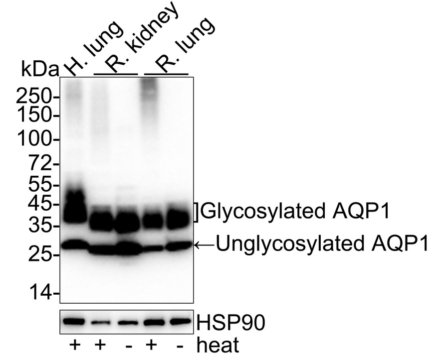
Western blot analysis of AQP1 on different lysates with Rabbit anti-AQP1 antibody at 1/2,000 dilution. Lane 1: Human lung tissue lysate, Lane 2: Rat kidney tissue lysate, Lane 3: Rat kidney tissue lysate (no heat), Lane 4: Rat lung tissue lysate, Lane 5: Rat lung tissue lysate (no heat), Notice: no heat means the lysate is not boiled. Lysates/proteins at 20 µg/Lane. Exposure time: 6 seconds; 4-20% SDS-PAGE gel. Proteins were transferred to a PVDF membrane and blocked with 5% NFDM/TBST for 1 hour at room temperature. The primary antibody at 1/2,000 dilution was used in 5% NFDM/TBST at 4℃ overnight. Goat Anti-Rabbit IgG - HRP Secondary Antibody at 1/50,000 dilution was used for 1 hour at room temperature.WB
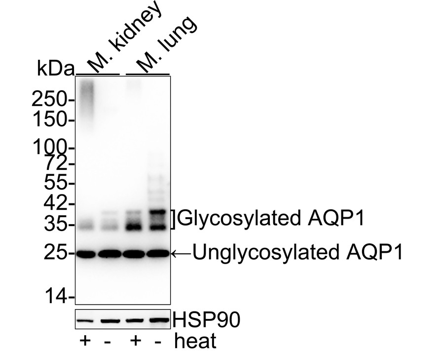
Western blot analysis of AQP1 on different lysates with Rabbit anti-AQP1 antibody at 1/5,000 dilution. Lane 1: Mouse kidney tissue lysate, Lane 2: Mouse kidney tissue lysate (no heat), Lane 3: Mouse lung tissue lysate, Lane 4: Mouse lung tissue lysate (no heat), Notice: no heat means the lysate is not boiled. Lysates/proteins at 20 µg/Lane. Exposure time: 24 seconds; Proteins were transferred to a PVDF membrane and blocked with 5% NFDM/TBST for 1 hour at room temperature. The primary antibody at 1/5,000 dilution was used in 5% NFDM/TBST at 4℃ overnight. Goat Anti-Rabbit IgG - HRP Secondary Antibody at 1/50,000 dilution was used for 1 hour at room temperature.ICC/IF
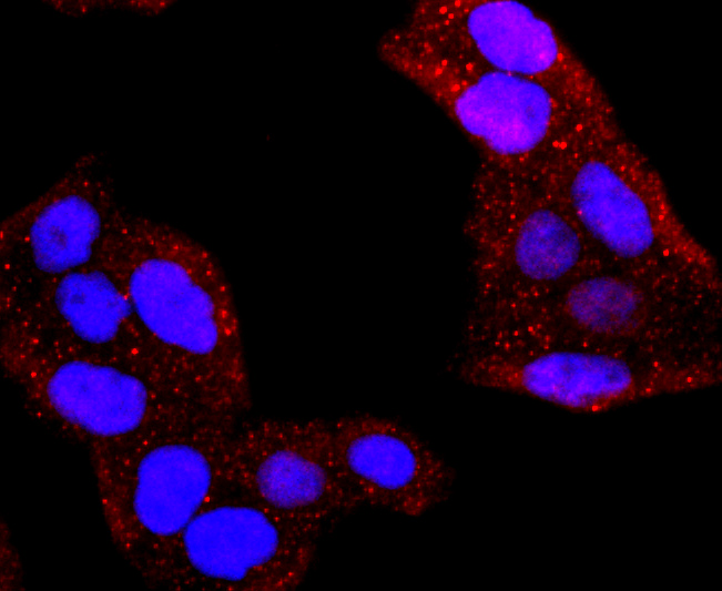
staining AQP1 in Hela cells (red). The nuclear counter stain is DAPI (blue). Cells were fixed in paraformaldehyde, permeabilised with 0.25% Triton X100/PBS.ICC/IF
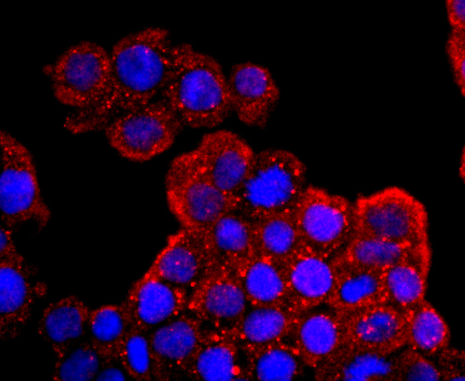
ICC staining AQP1 in SW480 cells (red). The nuclear counter stain is DAPI (blue). Cells were fixed in paraformaldehyde, permeabilised with 0.25% Triton X100/PBS.IHC
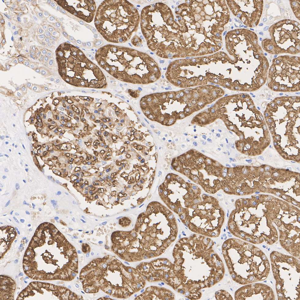
Immunohistochemical analysis of paraffin-embedded human kidney tissue with Rabbit anti-AQP1 antibody at 1/5,000 dilution. The section was pre-treated using heat mediated antigen retrieval with Tris-EDTA buffer (pH 9.0) for 20 minutes. The tissues were blocked in 1% BSA for 20 minutes at room temperature, washed with ddH2O and PBS, and then probed with the primary antibody at 1/5,000 dilution for 1 hour at room temperature. The detection was performed using an HRP conjugated compact polymer system. DAB was used as the chromogen. Tissues were counterstained with hematoxylin and mounted with DPX.IHC
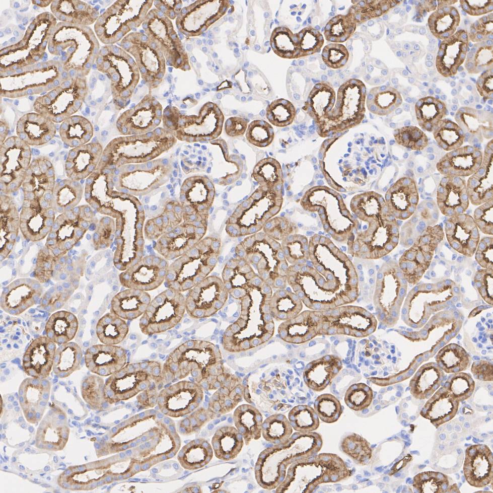
Immunohistochemical analysis of paraffin-embedded mouse kidney tissue with Rabbit anti-AQP1 antibody at 1/5,000 dilution. The section was pre-treated using heat mediated antigen retrieval with Tris-EDTA buffer (pH 9.0) for 20 minutes. The tissues were blocked in 1% BSA for 20 minutes at room temperature, washed with ddH2O and PBS, and then probed with the primary antibody at 1/5,000 dilution for 1 hour at room temperature. The detection was performed using an HRP conjugated compact polymer system. DAB was used as the chromogen. Tissues were counterstained with hematoxylin and mounted with DPX.IHC
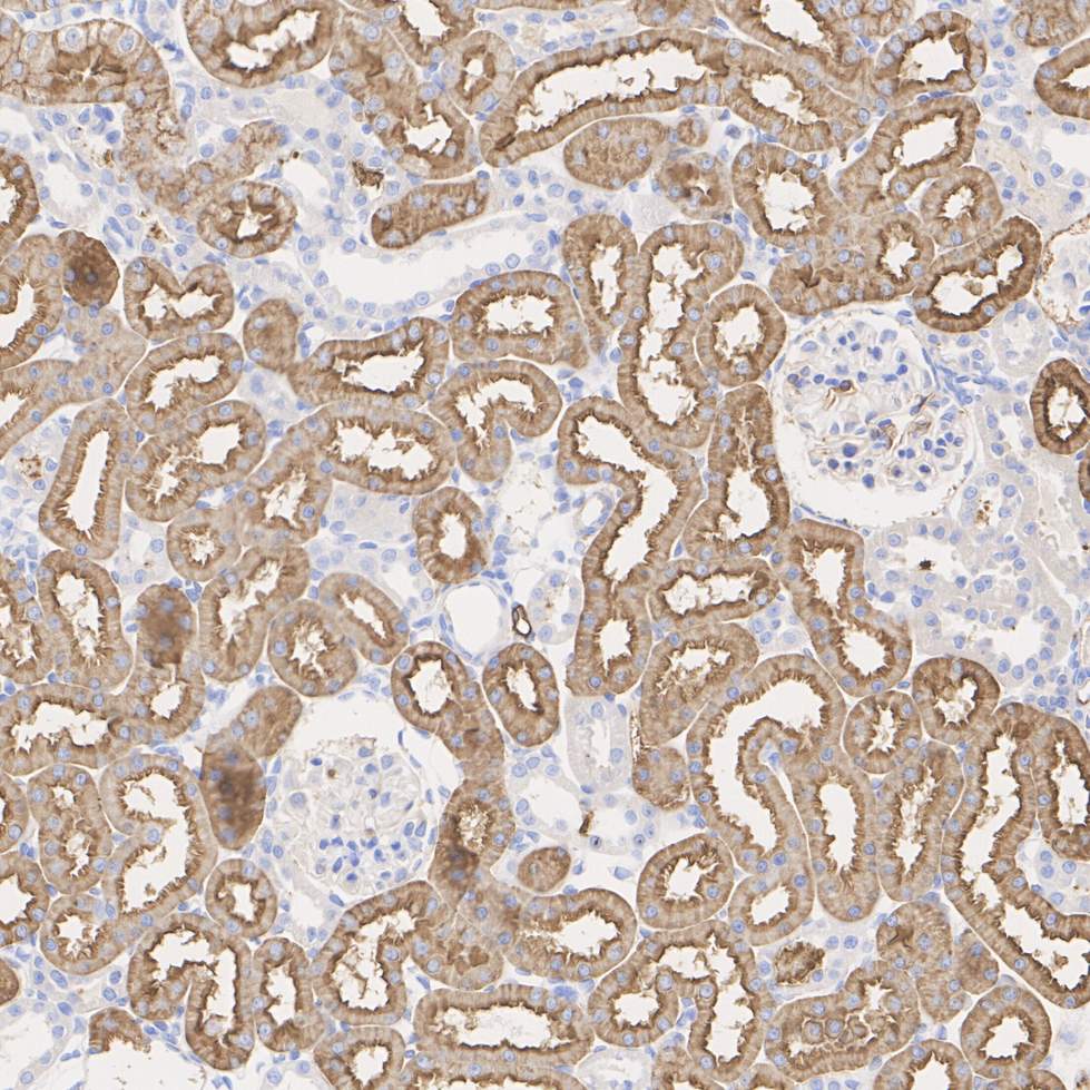
Immunohistochemical analysis of paraffin-embedded rat kidney tissue with Rabbit anti-AQP1 antibody at 1/5,000 dilution. The section was pre-treated using heat mediated antigen retrieval with Tris-EDTA buffer (pH 9.0) for 20 minutes. The tissues were blocked in 1% BSA for 20 minutes at room temperature, washed with ddH2O and PBS, and then probed with the primary antibody at 1/5,000 dilution for 1 hour at room temperature. The detection was performed using an HRP conjugated compact polymer system. DAB was used as the chromogen. Tissues were counterstained with hematoxylin and mounted with DPX.mIHC
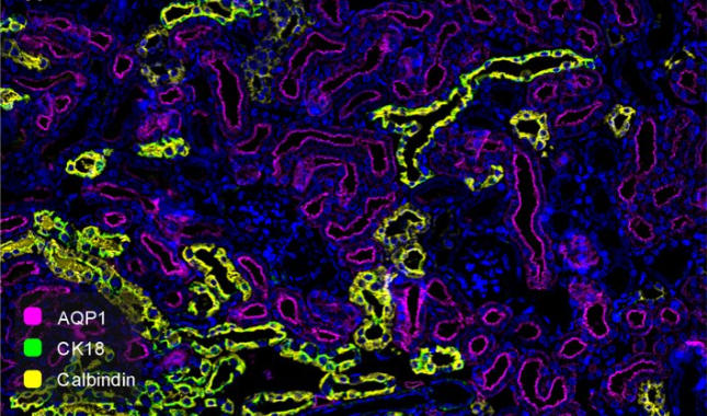
Fluorescence multiplex immunohistochemical analysis of mouse kidney (Formalin/PFA-fixed paraffin-embedded sections). Panel A: the merged image of anti-AQP1 (Violet), anti-CK18 (Green) and anti-Calbindin (Yellow) on kidney. HRP Conjugated UltraPolymer Goat Polyclonal Antibody was used as a secondary antibody. The immunostaining was performed with the Sequential Immuno-staining Kit. The section was incubated in three rounds of staining: in the order of anti-AQP1 (1/5,000 dilution),anti-CK18 (1/3,000 dilution) and anti-Calbindin (1/4,000 dilution) for 20 mins at room temperature. Each round was followed by a separate fluorescent tyramide signal amplification system. Heat mediated antigen retrieval with Tris-EDTA buffer (pH 9.0) for 30 mins at 95℃. DAPI (blue) was used as a nuclear counter stain. Image acquisition was performed with Zeiss Observer 7 Inverted Fluorescence Microscope.| Product Name | AQP1 Recombinant Rabbit Monoclonal Antibody |
|---|---|
| Antibody Type | Primary Antibodies |
| Product description | Aquaporins (AQPs) are a large family of integral membrane water transport channel proteins that facilitate the transport of water through the cell membrane. This function is conserved in animals, plants and bacteria. Many isoforms of Aquaporin have been identified in mammals, designated AQP0 through AQP10. Aquaporins are widely distributed and it is not uncommon for more than one type of AQP to be present in the same cell. Although most Aquaporins are only permeable to water, AQP3, AQP7, AQP9 and one of the two AQP10 transcripts are also permeable to urea and glycerol. AQP2 is the only water channel that is activated by vasopressin to enhance water reabsorption in the kidney collecting duct. Aquaporins are involved in renal water absorption, generation of pulmonary secretions, lacrimation and the secretion and reabsorption of cerebrospinal fluid and aqueous humor. AQP1 is an integral membrane protein expressed in erythrocytes and renal tubule cells. |
| Immunogen | Synthetic peptide within Human AQP1 aa 245-269 / 269. |
| Clonality | monoclonal |
|---|---|
| Isotype | IgG |
| Host Species | Recombinant rabbit |
| Tested Applications | ICC/IFIHCWBmIHC |
| WB:1:2000-1:5000 ICC/IF:1:500-1:2000 IHC:1:5000 mIHC:1:5000 |
|
| Species Reactivity | HumanMouseRat |
| Concentration | 1mg/ml |
| Purification | Protein A |
| Alternative Names | AQP 1 antibody
AQP CHIP antibody
AQP-1 antibody
AQP1 antibody
AQP1_HUMAN antibody
aquaporin 1 (channel-forming integral protein 28kDa CO blood group) antibody aquaporin 1 (Colton blood group) antibody Aquaporin CHIP antibody Aquaporin-1 antibody Aquaporin-CHIP antibody Aquaporin1 antibody Channel forming integral protein 28kDa antibody Channel like integral membrane protein 28 kDa antibody CHIP 28 antibody CHIP28 antibody CO antibody Colton blood group antibody Growth factor induced delayed early response protein antibody MGC26324 antibody Urine water channel antibody Water channel protein CHIP 29 antibody Water channel protein CHIP29 antibody Water channel protein for red blood cells and kidney proximal tubule antibody |
|---|---|
| Molecular Weight(MW) | 28kDa(Observed band size: 28~40kDa) |
| Cellular Localization | Cell membrane. |
WB
Western blot analysis of AQP1 on different lysates with Rabbit anti-AQP1 antibody at 1/2,000 dilution. Lane 1: Human lung tissue lysate, Lane 2: Rat kidney tissue lysate, Lane 3: Rat kidney tissue lysate (no heat), Lane 4: Rat lung tissue lysate, Lane 5: Rat lung tissue lysate (no heat), Notice: no heat means the lysate is not boiled. Lysates/proteins at 20 µg/Lane. Exposure time: 6 seconds; 4-20% SDS-PAGE gel. Proteins were transferred to a PVDF membrane and blocked with 5% NFDM/TBST for 1 hour at room temperature. The primary antibody at 1/2,000 dilution was used in 5% NFDM/TBST at 4℃ overnight. Goat Anti-Rabbit IgG - HRP Secondary Antibody at 1/50,000 dilution was used for 1 hour at room temperature.
WB
Western blot analysis of AQP1 on different lysates with Rabbit anti-AQP1 antibody at 1/5,000 dilution. Lane 1: Mouse kidney tissue lysate, Lane 2: Mouse kidney tissue lysate (no heat), Lane 3: Mouse lung tissue lysate, Lane 4: Mouse lung tissue lysate (no heat), Notice: no heat means the lysate is not boiled. Lysates/proteins at 20 µg/Lane. Exposure time: 24 seconds; Proteins were transferred to a PVDF membrane and blocked with 5% NFDM/TBST for 1 hour at room temperature. The primary antibody at 1/5,000 dilution was used in 5% NFDM/TBST at 4℃ overnight. Goat Anti-Rabbit IgG - HRP Secondary Antibody at 1/50,000 dilution was used for 1 hour at room temperature.
ICC/IF
staining AQP1 in Hela cells (red). The nuclear counter stain is DAPI (blue). Cells were fixed in paraformaldehyde, permeabilised with 0.25% Triton X100/PBS.
ICC/IF
ICC staining AQP1 in SW480 cells (red). The nuclear counter stain is DAPI (blue). Cells were fixed in paraformaldehyde, permeabilised with 0.25% Triton X100/PBS.
IHC
Immunohistochemical analysis of paraffin-embedded human kidney tissue with Rabbit anti-AQP1 antibody at 1/5,000 dilution. The section was pre-treated using heat mediated antigen retrieval with Tris-EDTA buffer (pH 9.0) for 20 minutes. The tissues were blocked in 1% BSA for 20 minutes at room temperature, washed with ddH2O and PBS, and then probed with the primary antibody at 1/5,000 dilution for 1 hour at room temperature. The detection was performed using an HRP conjugated compact polymer system. DAB was used as the chromogen. Tissues were counterstained with hematoxylin and mounted with DPX.
IHC
Immunohistochemical analysis of paraffin-embedded mouse kidney tissue with Rabbit anti-AQP1 antibody at 1/5,000 dilution. The section was pre-treated using heat mediated antigen retrieval with Tris-EDTA buffer (pH 9.0) for 20 minutes. The tissues were blocked in 1% BSA for 20 minutes at room temperature, washed with ddH2O and PBS, and then probed with the primary antibody at 1/5,000 dilution for 1 hour at room temperature. The detection was performed using an HRP conjugated compact polymer system. DAB was used as the chromogen. Tissues were counterstained with hematoxylin and mounted with DPX.
IHC
Immunohistochemical analysis of paraffin-embedded rat kidney tissue with Rabbit anti-AQP1 antibody at 1/5,000 dilution. The section was pre-treated using heat mediated antigen retrieval with Tris-EDTA buffer (pH 9.0) for 20 minutes. The tissues were blocked in 1% BSA for 20 minutes at room temperature, washed with ddH2O and PBS, and then probed with the primary antibody at 1/5,000 dilution for 1 hour at room temperature. The detection was performed using an HRP conjugated compact polymer system. DAB was used as the chromogen. Tissues were counterstained with hematoxylin and mounted with DPX.
mIHC
Fluorescence multiplex immunohistochemical analysis of mouse kidney (Formalin/PFA-fixed paraffin-embedded sections). Panel A: the merged image of anti-AQP1 (Violet), anti-CK18 (Green) and anti-Calbindin (Yellow) on kidney. HRP Conjugated UltraPolymer Goat Polyclonal Antibody was used as a secondary antibody. The immunostaining was performed with the Sequential Immuno-staining Kit. The section was incubated in three rounds of staining: in the order of anti-AQP1 (1/5,000 dilution),anti-CK18 (1/3,000 dilution) and anti-Calbindin (1/4,000 dilution) for 20 mins at room temperature. Each round was followed by a separate fluorescent tyramide signal amplification system. Heat mediated antigen retrieval with Tris-EDTA buffer (pH 9.0) for 30 mins at 95℃. DAPI (blue) was used as a nuclear counter stain. Image acquisition was performed with Zeiss Observer 7 Inverted Fluorescence Microscope.| Application Notes | WB:1:2000-1:5000 ICC/IF:1:500-1:2000 IHC:1:5000 mIHC:1:5000 |
|---|
| Form | Liquid |
|---|---|
| Storage Instructions | Store at +4℃ after thawing. Aliquot store at -20℃ or -80℃. Avoid repeated freeze / thaw cycles. |
| Storage Buffer | 1*TBS (pH7.4), 1%BSA, 40%Glycerol. Preservative: 0.05% Sodium Azide. |


